

On this page
When you study prokaryotic cells, one structure stands out: the mesosome. It is described as a specialised, differentiated form of the cell membrane. But the story of the mesosome is also one of biology's most famous cautionary tales. What started as a major discovery about bacterial respiration turned out to be an illusion created by the microscopes used to study it.
What is a mesosome in bacteria?
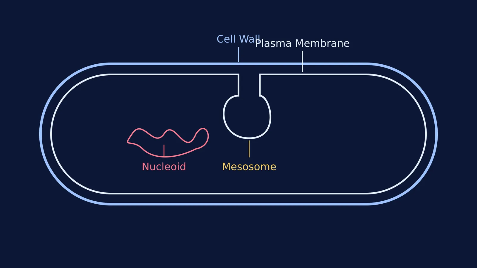
A mesosome is formed by the extensions of the plasma membrane into the bacterial cell. These extensions can take the form of vesicles, tubules, and lamellae.
Because prokaryotes lack membrane-bound organelles like mitochondria or a true nucleus, early biologists believed these complex infoldings were the cell's way of organising its internal machinery. They appeared as tangled, folded pockets on electron micrographs, seemingly anchored to key parts of the cell interior.
What are its functions?
If you are preparing for board exams or NEET, you must follow the official curriculum. The National Council of Educational Research and Training (NCERT) lists several vital functions for the mesosome. According to the textbook, mesosomes perform the following roles:
Cell wall formation: They are thought to synthesize and secrete the components needed to build the peptidoglycan cell wall.
DNA replication and distribution: During binary fission, the bacterial chromosome must be copied and split exactly in half. Mesosomes were believed to anchor the DNA, helping to pull the replicated chromosomes apart into the two daughter cells.
Respiration: Because bacteria lack mitochondria, the enzymes for aerobic respiration are located on the plasma membrane. The folded mesosome was seen as a way to increase the surface area for these respiratory enzymes, acting much like the cristae (inner folds) of a mitochondrion.
Secretion processes: They aid in the export of enzymes and other substances out of the cell.
Surface area enhancement: Overall, the infoldings simply increase the surface area of the plasma membrane and its enzymatic content, boosting the efficiency of cellular metabolism.
If you get stuck on the differences between prokaryotes and eukaryotes, Fermi's AI tutor can work out what you already know, build a plan for the session, and teach through it.
Are mesosomes real or just an artifact?
In the 1970s and 1980s, the scientific consensus on mesosomes flipped. Researchers realised that mesosomes were only visible when bacteria were prepared for electron microscopy using harsh chemical fixatives like osmium tetroxide or glutaraldehyde.
Chemical fixation is a slow process. As the chemicals seep into the cell to preserve it, they damage the bacterial plasma membrane. The stress causes the membrane to buckle and fold inwards, creating artificial pockets.
In a landmark 1981 paper, researchers H. R. Ebersold, J. L. Cordier and P. Lüthy tested this by using cryofixation. They froze the bacteria instantly using liquid nitrogen, stopping all biological processes in milliseconds before the membrane could distort. Under cryofixation, the mesosomes disappeared completely Archives of Microbiology. The researchers concluded that mesosomes are method-dependent artifacts.
The "mesosome story" is frequently taught in university biology as a lesson in methodological skepticism: just because a structure is visible and reproducible under a microscope does not mean it exists in a living cell. As imaging techniques evolved from slow chemical baths to rapid freezing, biologists had to unlearn decades of theories about how bacteria breathe and divide.
What are chromatophores in cyanobacteria?
While the mesosome turned out to be a structural artifact, not all membrane infoldings in bacteria are illusions. In cyanobacteria (blue-green algae), the plasma membrane folds inwards to form structures called chromatophores.
Unlike artifactual mesosomes, chromatophores are real, functional extensions of the membrane. They contain photosynthetic pigments, such as chlorophyll *a*, which allow cyanobacteria to capture light energy and carry out photosynthesis. Because prokaryotes do not have chloroplasts, these folded chromatophores provide the necessary surface area to hold the cell's light-harvesting machinery.
If you are comparing prokaryotic and eukaryotic machinery, you might also look at how eukaryotes divide transcription tasks among Three RNA polymerases in eukaryotes. You can read more about how other distinct structures arose in Viruses, viroids and lichens, the misfits of five-kingdom biology.
When you are ready to test your knowledge of the cell unit, the Fermi Problem Library has worked problems with a step-by-step solver that explains the reasoning rather than printing an answer.


