

On this page
Look at any cross-section of the human heart, and you will notice that it is not perfectly symmetrical. The muscular wall of the left ventricle is massively built, while the right ventricle looks like a thin crescent attached to its side. This is a classic biology question—and a frequent exam topic—but the reason comes down to basic physics and plumbing.
Why does the left ventricle pump harder?
The left ventricle is the heavy lifter of the circulatory system. Its job is to push oxygen-rich blood through the aorta and into the systemic circuit, reaching every tissue from the top of your head to the tips of your toes [[1]](https://www.ncbi.nlm.nih.gov/books/NBK470256/).
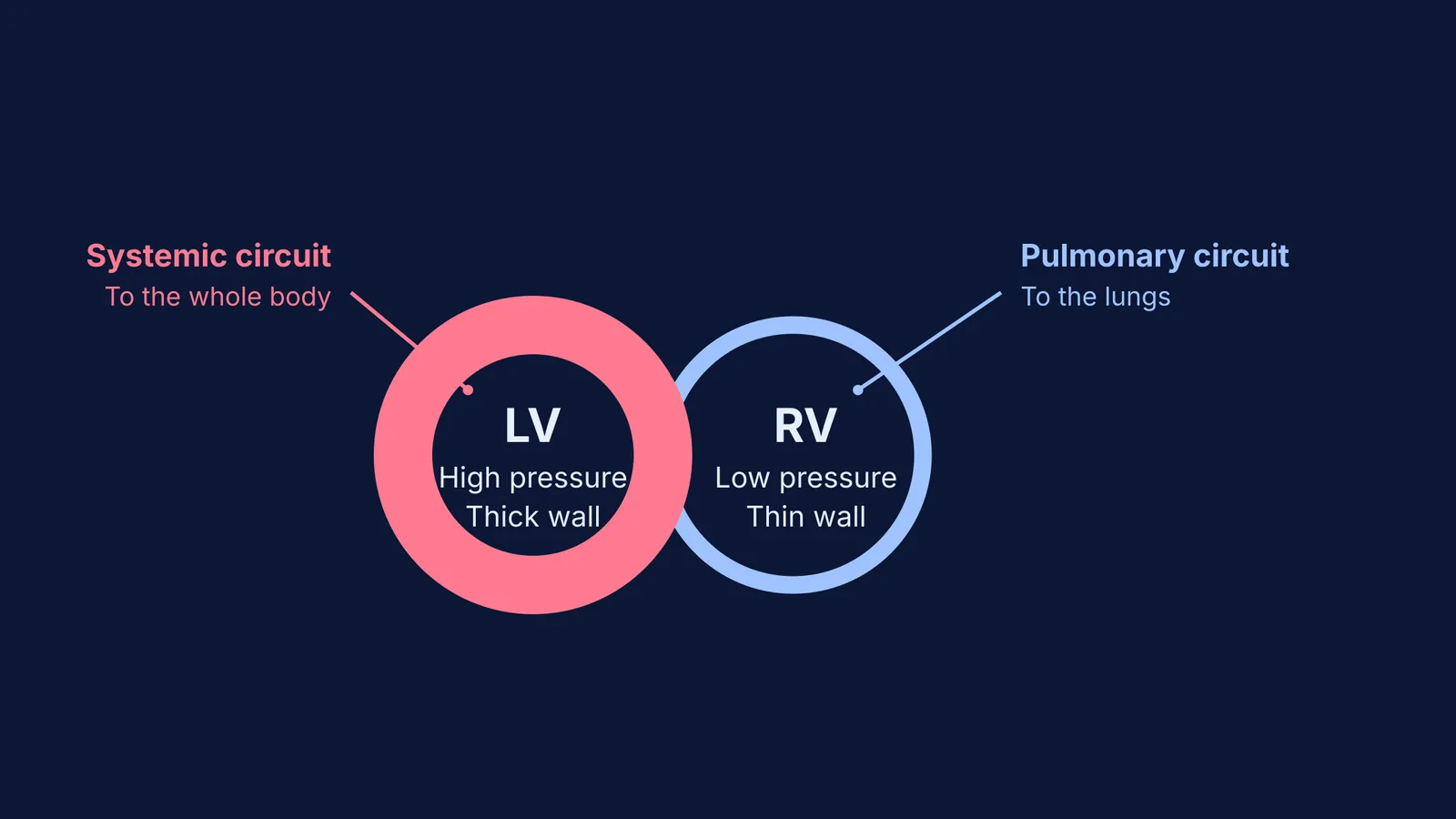
To drive blood through this massive network of vessels, it has to overcome high resistance. A healthy adult's left ventricle generates a high peak systolic pressure to keep blood flowing. To handle this intense workload, the left ventricle has a very thick muscular wall [[1]](https://www.ncbi.nlm.nih.gov/books/NBK470256/). Like any muscle that consistently lifts heavy weights, it is built for power.
Why is the right ventricle wall thinner?
The right ventricle handles the pulmonary circuit [[1]](https://www.ncbi.nlm.nih.gov/books/NBK470256/). As physician Ibn al-Nafis first described in the 13th century, blood from the right side of the heart travels only to the lungs to pick up oxygen.
Because the lungs sit right next to the heart in the chest cavity, the distance is short, and the resistance is low. More importantly, the delicate capillaries in the lungs would be damaged by high pressure. The right ventricle only needs to generate a much lower peak systolic pressure. Because the workload is significantly lower than the left side, the right ventricle's wall is much thinner [[1]](https://www.ncbi.nlm.nih.gov/books/NBK470256/).
How do the heart valves support this pressure?
To make sure blood flows in only one direction despite these immense forces, the ventricles rely on a system of one-way valves. The mitral valve sits between the left atrium and the left ventricle, while the aortic valve guards the exit to the aorta [[1]](https://www.ncbi.nlm.nih.gov/books/NBK470256/). When the massive left ventricle contracts, the mitral valve slams shut, preventing blood from flowing backwards. The high pressure forces the aortic valve open, sending blood to the body.
On the right side, the tricuspid valve separates the right atrium from the right ventricle, and the pulmonary valve separates the right ventricle from the pulmonary artery [[1]](https://www.ncbi.nlm.nih.gov/books/NBK470256/). Because the right ventricle operates at a much lower pressure, these valves do not endure the same extreme mechanical stress as the mitral and aortic valves, but their role in ensuring unidirectional flow to the lungs is just as vital.
How does the thick heart muscle get its blood?
You might think the heart absorbs oxygen directly from the blood inside its chambers, but the muscular walls—especially the thick left ventricle—require their own dedicated blood supply. The heart is fed by two main coronary arteries [[1]](https://www.ncbi.nlm.nih.gov/books/NBK470256/).
The left main coronary artery is particularly crucial because it carries most of the blood flow to the heart muscle. It branches into the left anterior descending artery and the circumflex artery, which together supply the massive left ventricular wall and the interventricular septum [[1]](https://www.ncbi.nlm.nih.gov/books/NBK470256/). Meanwhile, the right coronary artery primarily supplies the right atrium, the thinner right ventricle, and the inferior wall of the left ventricle. This extensive network ensures that the heart's heavy lifter never runs out of fuel.
How does this show up in diagrams?
If you are asked to label an unmarked diagram of a heart in a biology exam, the wall thickness is your best clue. The left ventricle will always have a thick, circular ring of muscle, often forming the bulk of the interventricular septum (the wall between the chambers). The right ventricle will usually appear as a thinner pouch draped over the right side.
Understanding these pressure differences also explains how the heart's electrical signals travel. The atrioventricular (AV) node and bundle branches have to distribute the signal so that this massive muscle contracts in a coordinated squeeze.
Why does structure follow function?
When you study the heart, it is easy to get lost in a list of valves and veins. But in biology, structure always follows function. High pressure demands thick walls; low pressure allows for thin ones. When surgeons like Christiaan Barnard performed the first human heart transplant, they were replacing a mechanical pump built precisely for these two very different jobs.
If you are untangling cardiovascular physiology, try exploring the Concept Library. It connects the anatomy of the heart to the physics of fluid pressure, and Fermi's AI tutor can walk you through the systemic and pulmonary circuits step by step until the whole map clicks into place.


