

On this page
When you study the human excretory system, mastering the nephron's loop of Henle and collecting duct is only half the battle. Chapter 19 of the NCERT Class 11 Biology textbook closes with three distinct disorders of the excretory system: uremia, renal calculi, and glomerulonephritis. Understanding where and why these occur is crucial for NEET and board exams, and for making sense of human physiology in a clinical setting. Rather than just memorising definitions, tracing the pathology back to the kidney's anatomy makes the concepts much easier to remember.
What is the difference between uremia, renal calculi and glomerulonephritis?
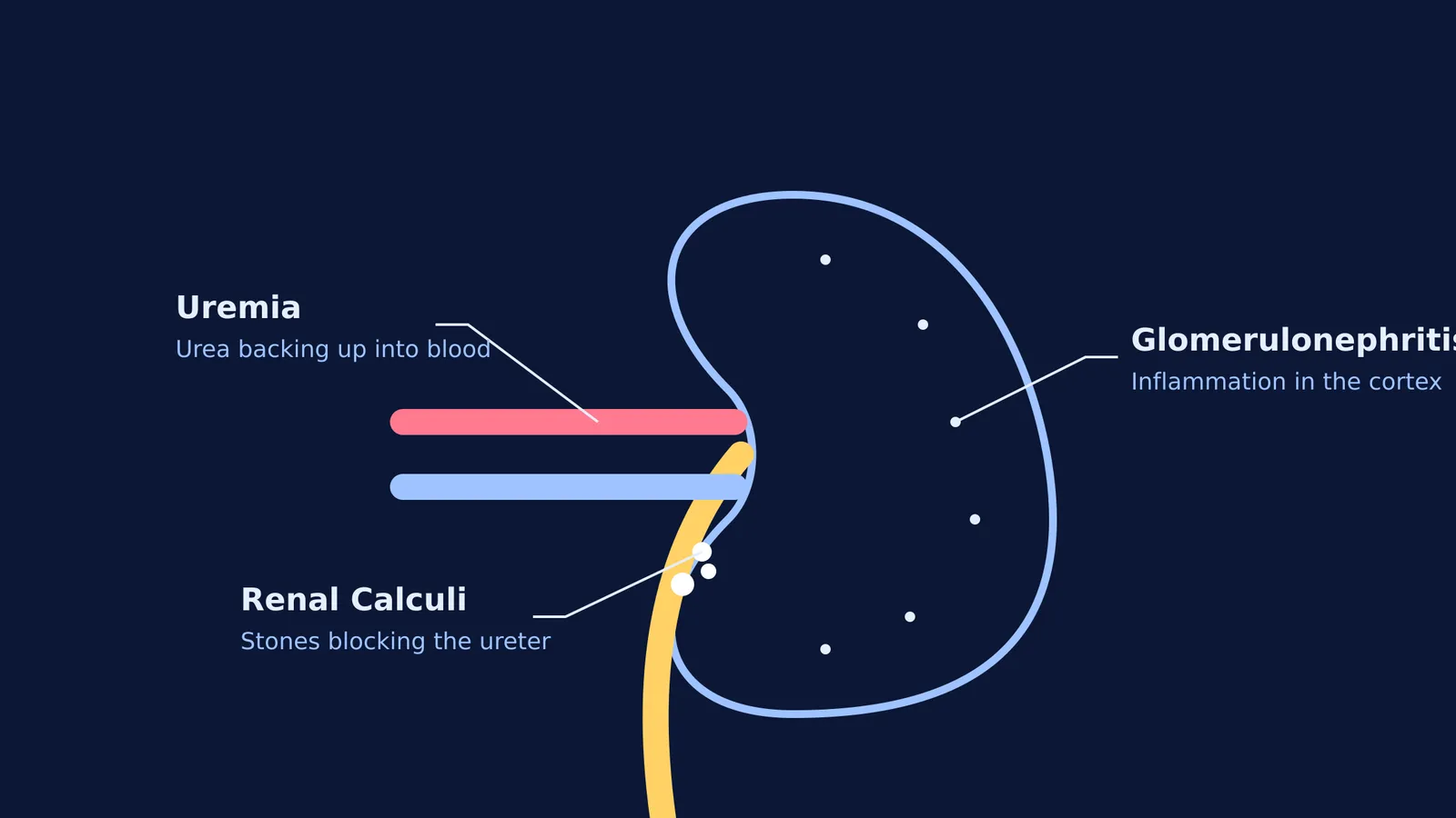
The easiest way to tell them apart is to look at where the physiological failure happens. Each disorder strikes a completely different part of the kidney's filtration, processing, and drainage system, leading to unique symptoms and treatments:
Glomerulonephritis is an issue with the filter itself (specifically the glomeruli located in the renal cortex).
Uremia is the systemic result of the filters failing entirely across the kidneys (toxins back up into the bloodstream).
Renal calculi are physical blockages in the drainage system (solid stones typically lodged in the renal pelvis or the ureter).
Uremia: why does urea accumulate in the blood?
Uremia literally translates to "urine in the blood." It is a severe clinical syndrome where the kidneys fail to clear nitrogenous waste, leading to a dangerous accumulation of urea and other toxins in the bloodstream. According to StatPearls, symptomatic uremia typically develops when the kidney's creatinine clearance drops below 10 to 20 mL/min.
Normally, your liver converts highly toxic ammonia into urea (a defining feature of ureotelic animals). This urea travels through the blood to the kidneys, where healthy nephrons filter it out into the urine. However, when glomerular filtration rates plummet due to chronic kidney disease, severe diabetes, or acute renal injury, that urea stays in the circulation. Because urea is harmful and highly toxic to the nervous system at elevated levels, severe uremia can lead to systemic organ failure, altered brain function, and eventually death if left untreated.
For patients suffering from severe uremia, the immediate and life-saving treatment is hemodialysis. An artificial kidney machine takes over the filtration process. Blood is routed through a dialyser, which uses a semi-permeable membrane and dialysate fluid to selectively remove urea and excess ions, restoring the electrolyte balance until a kidney transplant becomes possible.
Renal calculi: how do kidney stones form?
Renal calculi (commonly referred to as kidney stones) are an insoluble mass of crystallised salts formed within the kidney. While there are several types of stones, including uric acid and struvite stones, calcium oxalate stones are by far the most common in clinical cases.
Oxalate is a naturally occurring metabolic end-product and is also found in many foods, such as spinach, beetroot, and nuts. When oxalate levels in the urine are high and the overall urine volume is low (often due to dehydration), oxalate binds with calcium to form solid, pebble-like crystals. These crystals aggregate in the renal pelvis or ureter. As they grow or attempt to pass through the narrow ureter, they cause severe, tearing pain and block the flow of urine.
To prevent stone formation, the National Institute of Diabetes and Digestive and Kidney Diseases (NIDDK) notes that health professionals generally recommend drinking six to eight 8-ounce glasses of water a day (about 2 litres) to sufficiently dilute the urine. Paradoxically, reducing dietary calcium does not help; in fact, consuming calcium-rich foods alongside oxalate-rich foods allows them to bind harmlessly in the stomach and intestines instead of in the kidneys.
Glomerulonephritis: what inflames the glomeruli?
Glomerulonephritis is the inflammation of the glomeruli of the kidney. The glomeruli are the microscopic, high-pressure tufts of capillaries housed within Bowman's capsule, where blood is initially filtered to form the primary urine.
When these tiny, delicate filters become inflamed—often due to an aberrant autoimmune response or following a streptococcal infection—they become abnormally leaky. According to the NIDDK, damaged glomeruli allow proteins and sometimes red blood cells to leak into your urine. Instead of just letting water, glucose, and small metabolic waste molecules through, you lose essential formed elements of blood that should have remained safely in circulation. This disruption directly leads to proteinuria (excess protein in the urine) and hematuria (blood in the urine), alongside secondary symptoms like systemic swelling (edema) and high blood pressure.
Because the anatomical filters are compromised, untreated glomerulonephritis can eventually progress to chronic kidney disease. Over time, the loss of functioning nephrons leads right back to a severe drop in the filtration rate, ultimately resulting in uremia.
If you are trying to map out how these biological processes connect, the Fermi Concept Library holds every concept in biology, each with its own lesson and its place in the map, so you can trace exactly how blood becomes urine and where the microscopic filters break down.


