On this page
The human sperm is a highly specialised motile cell stripped down to the bare essentials: genetic material and the engine to deliver it.
Here is how the head, middle piece and tail work together to reach and penetrate the egg.
What are the parts of the human sperm?
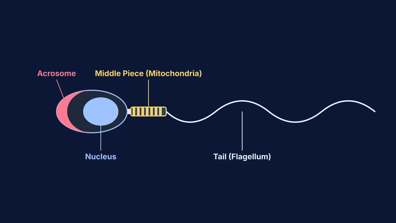
According to the WHO Laboratory Manual for the Examination and Processing of Human Semen (2021), a normal human spermatozoon measures approximately 50 to 60 µm in total length. It is divided into four main regions: the head, neck, middle piece and tail (flagellum).
To be considered morphologically normal, the head must be a smooth oval, the midpiece slender and axially attached, and the tail uncoiled.
What does the acrosome do in the sperm head?
The head contains a highly condensed haploid nucleus carrying the 23 paternal chromosomes. Covering the anterior half to two-thirds of the nucleus is a cap-like organelle called the acrosome.
The acrosome is derived from the Golgi apparatus during sperm development. It acts as a specialized secretory vesicle filled with hydrolytic enzymes, primarily hyaluronidase and acrosin. These enzymes have specific targets: hyaluronidase breaks down the hyaluronic acid holding together the corona radiata (the outer layer of follicular cells surrounding the egg), while acrosin digests the glycoproteins of the zona pellucida beneath it.
When the sperm successfully binds to the egg's receptors, it undergoes the acrosome reaction. The acrosomal membrane fuses with the sperm's plasma membrane, releasing these enzymes to dissolve a direct path to the egg's plasma membrane, allowing the genetic material to enter.
Why are mitochondria in the middle piece?
The middle piece connects the neck to the tail and serves as the cell's metabolic power plant. According to the WHO manual, the middle piece measures about 3.3 to 5.2 µm long, which is roughly the same length as the head.
Inside the midpiece, 50 to 75 mitochondria are wrapped end-to-end in a tight, helical sheath around the central axoneme core. This spiral arrangement is highly efficient, packing the maximum number of mitochondria into a confined space without increasing the sperm's width, which would create drag.
These mitochondria generate ATP via oxidative phosphorylation, using sugars like fructose found in seminal fluid. This ATP diffuses down the tail to fuel its whip-like beating. Interestingly, after fertilisation, the egg actively degrades these paternal mitochondria, which is why humans inherit mitochondrial DNA almost exclusively from their mothers.
Once the sperm successfully breaches the egg's barriers and fertilisation occurs, the sequence From fertilisation to implantation: the first week of pregnancy begins.
How does the tail drive motility?
The tail, or flagellum, is the longest segment of the sperm, extending approximately 45 µm (WHO). It is divided into a principal piece, which makes up most of its length, and a short end piece.
Inside the tail runs a central axoneme arranged in a classic 9+2 microtubule structure: nine peripheral doublet microtubules surrounding two central singlet microtubules. Attached to the peripheral doublets are dynein motor proteins. Using the ATP generated by the middle piece, these dynein arms "walk" along adjacent microtubules, causing them to slide past one another. Because the microtubules are anchored at the neck, this sliding translates into a bending motion, creating the complex wave-like beating that propels the sperm forward through the female reproductive tract.
If you need to sketch a sperm cell for a test, follow this four-step plan:
The head: Draw a flattened, paddle-like oval. Outline an inner cap over the front half and label it "Acrosome". Label the back half "Nucleus".
The middle piece: Add a short, thick cylinder below the head. Draw a zigzag or spiral line inside it and label it "Mitochondria".
The tail: Draw a long, tapering wave extending from the middle piece.
Function tags: Next to the labels, quickly note their roles: "Enzymes for penetration" for the acrosome, and "ATP for motility" for the mitochondria.
When you sit down to study, Fermi's AI tutor can help you connect these microscopic structures to their biological functions. Fermi teaches rather than answers: it holds a line of reasoning across a whole session. It works out what you already know and builds a plan for the session. You can also explore the Fermi Concept Library, which holds every concept in physics, chemistry, biology, mathematics and computing, each with its own lesson and its place in the map.
For a broader look at reproduction topics, review Parturition and lactation: the hormones behind birth and milk.