

On this page
If you are preparing for your Class 10 ICSE biology or physics exams—perhaps alongside your study of atmospheric refraction—drawing and labelling the human eye is a skill you will almost certainly need. The eye works like a highly sophisticated camera, capturing light, focusing it, and sending it to the brain as electrical signals.
The human eye is roughly spherical with a diameter of about 2.4 cm (Wikipedia). While the biological structures might seem complex at first, you can easily master them by breaking the eye down into its functional parts.
What are the parts of the human eye and their functions?
The front of the eye is designed to let light in and begin the focusing process.
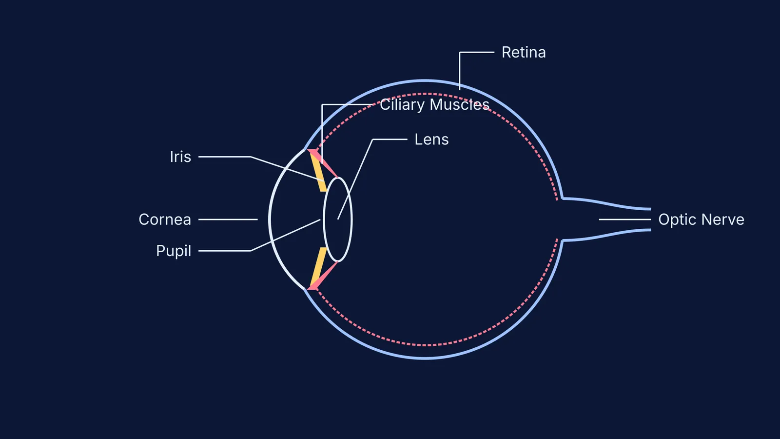
Cornea: This is the thin, transparent membrane bulging outwards on the front surface of the eyeball. The cornea is crucial because most of the refraction (bending) of light rays entering the eye occurs at its outer surface.
Iris: Located just behind the cornea, the iris is a dark, muscular diaphragm. It gives your eye its distinct colour (brown, blue, green). More importantly, the iris controls the size of the pupil.
Pupil: The pupil is the small black opening in the centre of the iris. Its main function is to regulate and control the amount of light entering the eye. In bright sunlight, the iris contracts the pupil to protect the inner eye; in a dark room, it expands the pupil to let in as much light as possible.
How does the ciliary muscle change the eye lens focus?
Once light passes through the pupil, it reaches the eye lens (or crystalline lens). This is a transparent, biconvex structure made of a fibrous, jelly-like material. Its job is to provide the finer adjustment of focal length needed to project a sharp, real and inverted image onto the back of the eye, applying the same principles of refraction you observe when tracing a ray through a glass slab.
The lens does not work alone. It is held in place and controlled by the ciliary muscles. These muscles modify the curvature of the eye lens to change its focal length—an ability known as accommodation. When the ciliary muscles relax, the lens becomes thinner, increasing its focal length to let you see distant objects clearly. When the muscles contract, the lens thickens, decreasing its focal length to focus on near objects, like a textbook.
Visualising how the ciliary muscles pull and relax the lens can be tricky on a static page. This is where tools like the Fermi Simulation Lab help, offering interactive simulations for the ideas that are easier to see than to read.
What is the function of the retina and optic nerve?
After the lens has focused the light, it projects an image onto the back wall of the eye.
Retina: This is a delicate, light-sensitive inner membrane that acts like a screen. According to Wikipedia, the retina contains an enormous number of light-sensitive cells called photoreceptors (rods and cones). When illuminated, these cells activate and convert the incoming light into electrical signals.
Optic Nerve: This nerve bundle acts as the eye's data cable. It transmits the electrical signals generated by the retina directly to the brain. The brain then interprets these signals and processes the information, allowing us to perceive objects upright and in three dimensions.
If you ever find yourself struggling to connect these biological functions to the physics of light, exploring the Fermi Concept Library can be incredibly useful. It holds every concept in physics, chemistry, biology, mathematics and computing, each with its own lesson and its place in the map, helping you tie topics like refraction and photoreception together.
How to label the eye for ICSE Class 10
Examiners look for precise placement of labels. Here is a practical framework to ensure you do not drop easy marks:
Draw the basic shape first: Start with a large circle for the sclera, but leave a gap at the front for the bulging cornea and a small gap at the back for the optic nerve.
Work front to back: Label the cornea on the very outside edge. Immediately behind it, draw the two flaps of the iris, and label the empty space between them as the pupil.
Place the lens and muscles: Draw the biconvex lens behind the iris, and connect its edges to the walls of the eye using small triangles to represent the ciliary muscles.
Mark the inner screen: Draw a dotted line along the inside back wall to represent the retina.
Draw the exit path: Label the bundle of fibres exiting the back gap as the optic nerve.
If you are stuck on exactly how much detail to include or how to answer specific application questions on this topic, Fermi's AI tutor can help. It holds a line of reasoning across a whole session, notices where a student is stuck, and decides whether to explain, ask, or show.
By learning what each part does rather than just memorising a diagram, you will find it much easier to tackle both straight recall questions and complex application problems in your board exams.


