

On this page
Why does the immune system attack a transplanted organ?
When a vital organ fails, transplantation offers a lifesaving medical remedy. To your immune system, however, a transplanted organ resembles an aggressive mass of foreign tissue. The human body has spent hundreds of millions of years refining molecular mechanisms to distinguish "self" from "non-self". The moment living cells from a genetically non-identical donor enter the body, that defensive apparatus mobilizes.
Transplantation biology established this immunological basis through Peter Medawar's experiments in 1944. Working with rabbit skin grafts, Medawar showed that autografts healed permanently, while homografts from unrelated donors were rejected within 8 to 12 days (Medawar, 1944). When he transplanted skin from that same donor a second time, the host destroyed it in just 4 to 6 days. This "second-set phenomenon" proved that rejection is an acquired immune response with memory and specificity.
In the NCERT Class 12 Biology syllabus, students learn that graft rejection is driven by cell-mediated immunity (CMI) mediated by T-lymphocytes. These cells mature in the thymus before circulating, a developmental milestone covered in our article on Jacques Miller and the role of the thymus in immunity.
How do T-lymphocytes distinguish self from non-self?
Every nucleated human cell displays surface proteins called Human Leukocyte Antigens (HLA), encoded by the Major Histocompatibility Complex (MHC) on chromosome 6. MHC Class I molecules (HLA-A, HLA-B, HLA-C) sit on all nucleated cells, presenting intracellular peptides to CD8+ cytotoxic T cells. MHC Class II molecules (HLA-DR, HLA-DQ, HLA-DP) appear on specialized antigen-presenting cells (dendritic cells, macrophages, B cells), presenting engulfed peptides to CD4+ helper T cells.
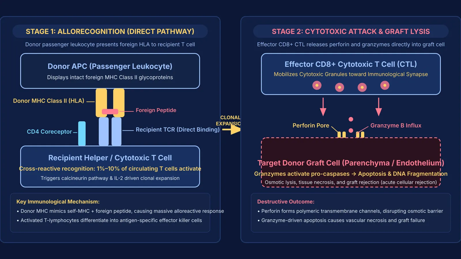
When an organ is transplanted, the recipient's T-cell receptors (TCRs) recognize the foreign HLA molecules through two distinct pathways (Ali et al., 2013):
Direct allorecognition: Recipient T cells bind intact foreign MHC on donor dendritic cells ("passenger leukocytes") that migrate from the graft. Because foreign MHC structures cross-react broadly with host receptors, between 1% and 10% of a recipient's entire circulating T-cell pool can respond immediately.
Indirect allorecognition: Recipient antigen-presenting cells process shed donor proteins and present donor peptides on self-MHC Class II molecules to CD4+ helper T cells, driving sustained chronic inflammation.
Once activated, cytotoxic T cells release perforin to puncture donor cell membranes and granzymes to trigger programmed cell death (apoptosis). If you are mapping the molecular steps between antigen presentation and cell-mediated cytotoxicity, the Fermi Concept Library covers every concept in physics, chemistry, biology, mathematics and computing, each with its own lesson and its place in the map.
Why do tissue matching and blood group matching matter?
To reduce the frequency of alloreactive T-cell clones, transplant teams perform HLA typing. The clinical standard prioritizes three gene loci: HLA-A, HLA-B, and HLA-DR. Because every individual inherits one set of alleles from each parent, there are six main antigens evaluated in a standard clinical match (a "six-antigen match").
Clinical outcomes demonstrate that matching directly prolongs graft survival. In an analysis of 133,442 deceased-donor kidney transplants from the Collaborative Transplant Study (Opelz & Döhler, 2007), zero-mismatched kidneys achieved approximately 17% higher 10-year graft survival than fully mismatched grafts. In India, allocation guidelines from the National Organ and Tissue Transplant Organisation (NOTTO) award scoring points for close HLA compatibility in deceased-donor kidney allocation.
Identical twins share identical MHC genes, allowing organs to survive indefinitely without immunosuppressive drugs. This was first demonstrated in 1954, as explored in our history of Joseph Murray and the first successful kidney transplant.
What happens during hyperacute, acute, and chronic rejection?
Graft rejection occurs across three distinct clinical timelines based on the underlying immunological mechanism (Nankivell & Alexander, 2010):
Hyperacute rejection (minutes to hours): Caused by pre-existing donor-specific antibodies (such as anti-ABO blood group antigens or anti-HLA antibodies from prior pregnancies, blood transfusions, or earlier grafts). These antibodies bind vascular endothelium, activate the complement cascade, cause thrombosis, and result in immediate graft ischemia.
Acute rejection (days to months): Primarily mediated by cell-mediated immunity. Recipient CD8+ T cells infiltrate parenchymal tissue, causing tubulitis in kidneys or endotheliitis in heart grafts. Acute rejection can also involve de novo antibody formation.
Chronic rejection (months to years): Characterized by progressive tissue remodeling, interstitial fibrosis, and vascular occlusive disease (transplant vasculopathy). It results from low-grade cellular infiltration and persistent donor-specific antibodies resistant to standard therapy.
How do doctors test compatibility before surgery?
Transplant immunology teams use a four-step laboratory checklist before clearing an organ for surgery:
Step 1 (ABO Grouping): Verifies standard red cell compatibility to prevent immediate endothelial antibody binding.
Step 2 (HLA Typing): Uses DNA sequencing to identify specific alleles across HLA-A, HLA-B, HLA-C, and HLA-DR loci.
Step 3 (Panel Reactive Antibody Screen): Measures the percentage of standard HLA variants against which the recipient carries pre-formed antibodies.
Step 4 (Direct Crossmatch): Mixes recipient serum with donor lymphocytes; a positive crossmatch detects pre-existing donor-specific antibodies and serves as an absolute surgical contraindication.
These immunological principles link directly to questions tested across board exams and competitive entrance tests, which we analyze further in our guide to Human health and disease for NEET: pathogens, immunity, vaccines. When practicing multi-step genetics or immunology problems, the Fermi Problem Library provides worked problems with a step-by-step solver that explains the reasoning rather than printing an answer.


