

On this page
What is the difference between free and bound ribosomes?
Structurally and biochemically, free ribosomes and membrane-bound ribosomes are identical. In eukaryotic cells, both are 80S ribonucleoprotein particles composed of a 60S large subunit and a 40S small subunit, as defined in the NCERT Class 11 Biology curriculum. They share a common cytosolic pool, separating into "free" and "bound" states that were first quantified through sedimentation analysis, which you can explore in our guide to ultracentrifugation and sedimentation coefficients. Subunits return to the cytoplasm after translation and reassemble arbitrarily for the next cycle.
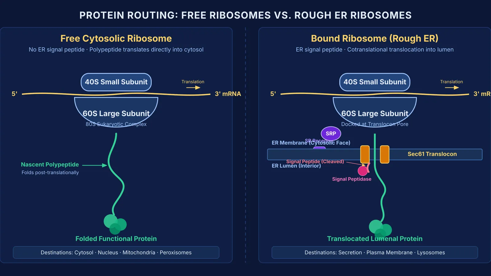
What differentiates them is the specific protein being manufactured. All translation begins on free ribosomes in the cytosol. If the messenger RNA encodes a polypeptide for internal cellular duties—such as enzymes that drive glycolysis—the ribosome remains freely suspended throughout synthesis, releasing the completed protein directly into the cytosol.
If the mRNA encodes a protein destined for secretion, insertion into membranes, or packaging within lysosomes, a specialized sequence at the front of the growing polypeptide directs the ribosome to dock against the cytosolic face of the rough endoplasmic reticulum (RER).
How does a signal peptide direct a ribosome to the ER?
The mechanism guiding ribosomes to the ER membrane is the signal hypothesis, demonstrated by Günter Blobel and Bernhard Dobberstein in their 1975 investigation published in the *Journal of Cell Biology*. Blobel later received the 1999 Nobel Prize in Physiology or Medicine for proving that proteins carry intrinsic signals that govern their cellular transport.
As established by Blobel and Dobberstein in their 1975 protein translocation study, the N-terminal signal peptide is typically 15 to 30 amino acids long, with a core of 6 to 15 hydrophobic residues. Routing unfolds in five steps:
Emerging sequence: Once the nascent chain reaches roughly 70 amino acids, having traversed the 30 to 40 amino acid ribosomal exit tunnel, the hydrophobic signal peptide emerges into the cytosol.
Recognition by SRP: The signal peptide binds the Signal Recognition Particle (SRP). SRP is a cytosolic ribonucleoprotein complex made of one 7SL RNA molecule of about 300 nucleotides and six polypeptide chains, shown by Peter Walter and Günter Blobel in the *Journal of Cell Biology* to arrest translation elongation until membrane docking occurs.
Elongation arrest: SRP binding pauses translation, preventing the polypeptide from prematurely folding or aggregating in the cytosol.
Membrane docking: The ribosome-SRP complex migrates to the rough ER membrane, where SRP binds its membrane-bound receptor, aligning the exit tunnel over the Sec61 translocon pore.
Translocation and cleavage: GTP hydrolysis by SRP and its receptor releases SRP into the cytosol. Translation resumes, feeding the polypeptide cotranslationally through the translocon into the ER lumen, where signal peptidase cleaves off the signal sequence.
Which proteins are made on free ribosomes versus the rough ER?
This cellular division of labor maintains compartmentalization, an architectural principle detailed in our guide to organelle functions in cell biology.
Free ribosomes synthesize proteins that function in non-secretory environments:
Cytosolic enzymes: Catalysts for metabolic pathways, including hexokinase, aldolase, and pyruvate kinase in glycolysis.
Cytoskeletal filaments: Structural proteins that provide cellular shape, including actin, tubulin, and intermediate filaments.
Nuclear proteins: Polypeptides with a Nuclear Localization Signal, including histones and DNA polymerases, imported through nuclear pores after translation.
Organellar proteins: Mitochondrial, chloroplast, and peroxisomal enzymes, such as catalase, imported post-translationally through membrane translocases.
Membrane-bound ribosomes synthesize proteins for the endomembrane system and secretion. George Palade and James Jamieson traced this pathway using a 3-minute pulse with radioactive amino acids in pancreatic cells, published in the *Journal of Cell Biology* and explored in our story on George Palade's pulse-chase experiments on the secretory pathway:
Secretory proteins: Hormones, digestive enzymes, and antibodies, including insulin and pepsinogen.
Integral membrane proteins: Transmembrane proteins with stop-transfer sequences, including ion channels, pumps, and cell-surface receptors.
Lysosomal hydrolases: Digestive enzymes such as proteases and acid phosphatase, tagged with mannose-6-phosphate in the Golgi.
How do you solve protein destination questions in NEET?
In NEET-UG and Class 11 exams, questions test where an enzyme or hormone is manufactured. A four-step check keeps them straight:
Identify the working site: Proteins operating outside the cell, within membranes, or in lysosomes are made on rough ER ribosomes.
Check for internal targets: Proteins operating in the cytosol, nucleus, mitochondria, or peroxisomes are made on free ribosomes.
Confirm timing: Rough ER proteins enter the lumen cotranslationally, while free ribosome proteins enter target organelles post-translationally.
Verify the pathway: Secreted and membrane proteins travel strictly from Rough ER to Golgi, then to vesicles or lysosomes.
Consider human insulin versus DNA polymerase. Insulin is secreted by pancreatic beta cells into blood, requiring synthesis on rough ER ribosomes, signal cleavage, and Golgi packaging. DNA polymerase acts in the nucleus replicating chromatin, requiring synthesis on free cytosolic ribosomes and nuclear pore import.
When practicing cellular pathways and organelle destinations, the Fermi Problem Library offers worked problems with a step-by-step solver that explains the reasoning rather than printing an answer. For students reviewing how the endomembrane system connects with cell biology as a whole, the Fermi Concept Library covers every concept in physics, chemistry, biology, mathematics and computing, each with its own lesson and its place in the map.


