

On this page
Understanding the development of the ovarian follicle is a core part of human reproduction. Whether you are preparing for a board exam or a competitive test like NEET, knowing exactly when specific structures appear—and what they do—can save you marks. Biology exams frequently test your ability to match a structure to its stage.
What are the stages from primary to Graafian follicle?
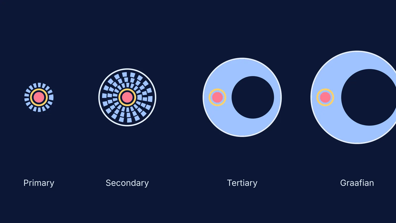
Folliculogenesis is the continuous process where a small, immature follicle grows and changes into a mature structure ready for ovulation. The process moves through four main stages: primary, secondary, tertiary (or antral), and Graafian (or preovulatory).
At each stage, the follicle gains new layers and functions. The defining changes involve the granulosa cells multiplying, the surrounding tissue organising into theca layers, and a fluid-filled cavity called the antrum opening up inside. Memorising these changes by drawing them is one of the best ways to prepare, just as it is for other NCERT biology diagrams for NEET.
When do the theca layers and zona pellucida form?
The earliest major changes happen in the primary and secondary stages.
The primary follicle is marked by the appearance of a glycoprotein capsule called the zona pellucida. This capsule forms a protective ring directly around the oocyte. The zona pellucida remains with the oocyte after ovulation. The surrounding cells also change shape from flat to cuboidal granulosa cells. At this stage, the follicle grows significantly, reaching almost 0.1 mm in diameter.
As it becomes a secondary follicle, new stroma-like tissue is recruited to form the theca layers. These cells organise into two distinct bands: the highly vascular theca interna, which eventually produces androgens, and the outer fibrous theca externa. A late-term secondary follicle typically features approximately nine layers of granulosa cells.
When does the antrum appear in a follicle?
The defining feature of the tertiary follicle is the formation of the follicular antrum. The antrum is a fluid-filled cavity rich in hyaluronic acid that begins to form in the late secondary stage and fully opens up in the tertiary stage.
Once the antrum forms, the granulosa cells differentiate into distinct regions. One such region is the cumulus oophorus, a cluster of cells that connects the membrana and corona radiata granulosa cells together. During a typical menstrual cycle, a cohort of 5 to 7 follicles is recruited, but only one survives to become the dominant Graafian follicle.
How does the Graafian follicle rupture during ovulation?
The Graafian follicle is the final preovulatory stage where the antrum reaches its maximum volume. During the follicular phase, the dominant follicle grows at a rate of about 1.5 mm per day. Ovulation typically occurs once the follicle reaches at least 14 mm in diameter, and it can grow up to 20 mm.
At the end of the follicular phase, a massive surge in luteinising hormone (LH) activates pro-inflammatory genes, causing the follicle wall to break. This creates a weakened, thinned area on the surface of the ovary known as the stigma. Finally, the wall ruptures at the stigma, gently expelling the oocyte—still surrounded by its zona pellucida and cumulus oophorus—into the fallopian tube. The remaining cells collapse and transform into the corpus luteum.
How to revise follicle stages for exams?
To avoid confusing the layers under pressure, use this active-learning checklist:
Draw from memory: Sketch the four stages side-by-side without looking at your book.
Label the first appearances: Mark exactly where the zona pellucida (primary), theca layers (secondary), and antrum (tertiary) first show up.
Trace the hormones: Note that the theca interna produces androgens, which the granulosa cells then convert into oestrogen.
Link to the wider system: Fit this process into your broader human physiology for NEET revision plan.
Understanding these structures also connects deeply with other biological principles, such as those covered in our guide to evolution for NEET.
If you are struggling to picture how these three-dimensional structures fit together, the Fermi Concept Library has every concept in physics, chemistry, biology, mathematics and computing, each with its own lesson and its place in the map. When you are ready to test your knowledge, the Fermi Problem Library has worked problems with a step-by-step solver that explains the reasoning rather than printing an answer.


