

On this page
Epithelial tissue provides a continuous, protective layer for the body and its organs (Wikipedia). Based on the number of cell layers, it divides into two major groups: simple epithelium and compound epithelium. Understanding where each type sits is a matter of linking the tissue's shape to the job it has to do in that organ.
What is simple squamous epithelium?
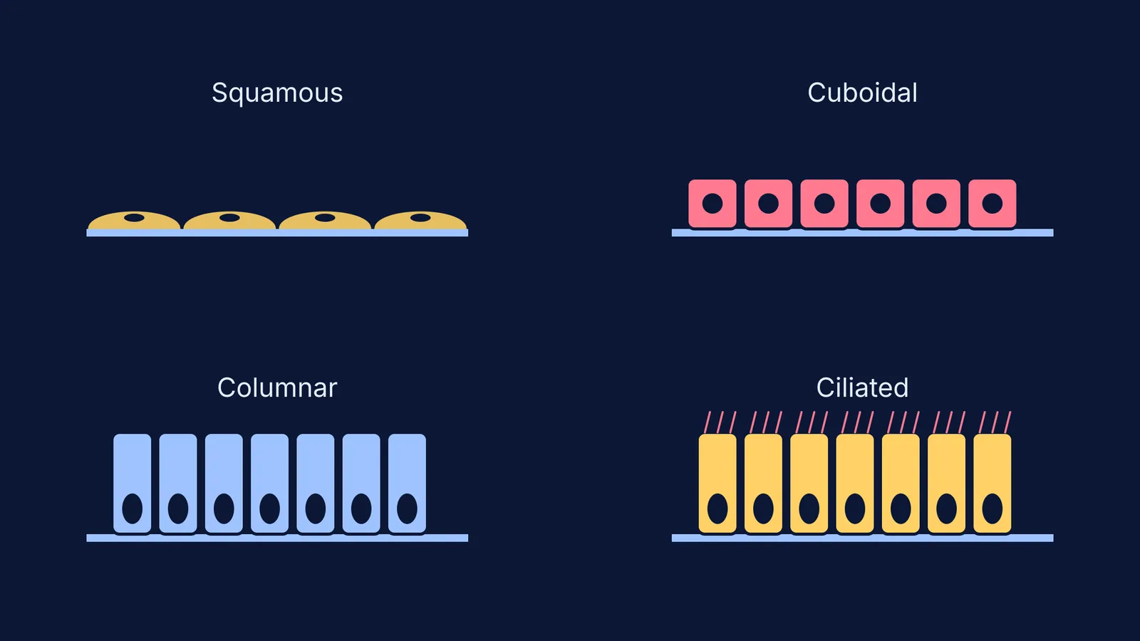
Squamous epithelium consists of a single, very thin layer of flattened cells with irregular boundaries (Wikipedia). Because it is so thin, it is perfectly built to form a diffusion boundary where gases, fluids, or ions need to pass through quickly without encountering resistance.
You will find simple squamous epithelium lining the walls of blood vessels (where it is known as the endothelium) and the air sacs (alveoli) of the lungs. For example, when you take a breath, oxygen moves from your lungs into the blood by passing straight through the squamous cells of the alveoli and the adjacent squamous cells of the capillary wall. This minimal barrier ensures that gas exchange happens rapidly across the membrane.
Where is simple cuboidal epithelium located?
Simple cuboidal epithelium is a single layer of cube-like cells. Because these cells have more volume than squamous cells, they house more organelles, making their main functions secretion and absorption rather than simple diffusion.
It is commonly found lining the ducts of small glands (such as the salivary glands) and in the tubular parts of nephrons in the kidneys during urine formation. A notable example is the proximal convoluted tubule (PCT) of the nephron. Here, the cuboidal epithelium is covered in microscopic finger-like projections called microvilli. This "brush border" massively increases the surface area available to reabsorb water, glucose, and essential ions back into the blood.
What does simple columnar epithelium do?
When cells need to secrete digestive juices or absorb nutrients on a larger scale, the body relies on simple columnar epithelium. This tissue is composed of a single layer of tall and slender cells, looking like standing columns, with their elongated nuclei located near the base rather than in the centre.
Simple columnar epithelium lines most of the digestive tract, including the stomach and the large and small intestines. In the stomach, these tall cells secrete mucus to protect the stomach lining from its own harsh acidic environment. In the intestine, just like the cuboidal cells in the kidney, they often feature microvilli on their free surface to maximise the absorption of digested food. Mixed in among these columnar cells are specialised goblet cells that produce protective mucus.
Where is ciliated epithelium located?
Sometimes, cuboidal or columnar cells bear tiny, motile, hair-like outgrowths on their free apical surface, known as cilia (Wikipedia). Their job is to move particles, fluids, or mucus steadily in a specific direction over the epithelium.
Ciliated epithelium is mainly present in the inner surface of hollow organs such as the small bronchioles of the respiratory tract and the fallopian tubes. In the respiratory tract, the beating cilia sweep dust-trapping mucus outward and away from the lungs. In the fallopian tubes, they act like a conveyor belt to help waft the ovum safely towards the uterus. The internal structure of these cilia shares the same complex 9 + 2 axoneme arrangement found in cilia and flagella throughout biology.
To explore how epithelial structures connect across organ systems, the Concept Library includes every concept in biology, each with its own lesson and its place in the map.
What is compound epithelium?
While simple epithelium is just one cell thick, compound (or stratified) epithelium is made of more than one layer of cells. Because of these multiple layers, it does not do much secretion or absorption; instead, its primary role is to provide robust protection against chemical and mechanical stresses.
Compound epithelium covers the dry surface of the skin (the epidermis), where it takes constant wear, tear, and dehydration. In this location, the outermost layers are dead and packed with keratin. It also covers moist surfaces that experience friction, such as the buccal cavity (the inside of the mouth), the pharynx, the inner lining of ducts of salivary glands, and pancreatic ducts.
Where is transitional epithelium found?
A unique type of compound epithelium is transitional epithelium, or urothelium (Wikipedia). This multi-layered tissue is designed to stretch and change shape as an organ fills with fluid. It is found lining the urinary bladder, ureters, and parts of the urethra, allowing the bladder to expand dramatically without tearing.
When working through practice questions on tissue classification, the Fermi Problem Library provides worked problems with a step-by-step solver that explains the reasoning rather than printing an answer. Understanding how these tissue layers function will also help you when you study the vessels that carry the formed elements of blood.


