

On this page
The cell is not a loose bag of water and organelles. Just as the human skeleton provides structural support, every eukaryotic cell relies on an internal, dynamic framework called the cytoskeleton.
In short: The cytoskeleton gives a cell its shape, anchors organelles, and allows for movement. According to OpenStax Biology, it relies on three structures: thick microtubules (25 nm), thin microfilaments (7 nm), and intermediate filaments (8–10 nm).
What does the cytoskeleton do?
A cell's cytoskeleton has three main responsibilities: mechanical support, motility, and the maintenance of shape. It is an elaborate network of filamentous proteinaceous structures spread throughout the cytoplasm.
Without this framework, cells would collapse under physical stress, and organelles would drift aimlessly. The cytoskeleton allows cells to hold their form against external pressure. It also directs the traffic inside the cell, making sure that vesicles, proteins, and chromosomes reach their exact destinations. If you find it hard to picture how all these parts interact during cell processes, the Fermi Concept Library contains a dedicated lesson for every biology concept and shows its place in the wider topic map.
Functions of microtubules: the transport network
Microtubules are the thickest of the cytoskeletal filaments. As detailed in OpenStax Biology, they are hollow cylinders approximately 25 nm in diameter, composed of the protein tubulin.
These structures act as the cell's rigid highways. Their primary functions include:
Intracellular transport: Motor proteins like kinesin and dynein "walk" along microtubules, hauling vesicles and organelles from the centre of the cell to the edges, or vice versa.
Cell division: During mitosis and meiosis, microtubules assemble into the spindle fibres. These fibres attach to chromosomes and pull them apart, ensuring each new daughter cell receives the correct genetic material.
Centrioles: In animal cells, the centrosome contains two cylindrical structures called centrioles. According to standard reference materials, each centriole is built from nine evenly spaced peripheral fibrils of tubulin arranged in a 9+0 pattern, forming a core hub connected by radial spokes.
Microfilaments and cell movement
Microfilaments are the thinnest components of the cytoskeleton. They measure about 7 nm across (OpenStax Biology) and are primarily composed of two intertwined strands of the globular protein actin.
While microtubules resist compression like steel poles, microfilaments bear tension like cables. Their functions are heavily tied to cellular shape changes and movement:
Amoeboid movement: Microfilaments allow cells, such as the white blood cells (the formed elements of blood responsible for immunity), to crawl. They push the cell membrane forward to form pseudopodia (false feet).
Muscle contraction: In muscle tissue, actin microfilaments slide past another protein called myosin, causing the entire muscle cell to contract and generate force.
Cytokinesis: When an animal cell finishes dividing its nucleus, a contractile ring of actin microfilaments pinches the cytoplasm inwards. This creates a cleavage furrow that eventually splits the cell into two distinct daughter cells.
Cytoplasmic streaming: In plant cells, microfilaments drive cyclosis. This is a circular flow of cytoplasm that helps distribute nutrients, enzymes, and chloroplasts evenly around the large central vacuole.
Intermediate filaments: anchoring the cell
Sitting between the thick microtubules and thin microfilaments are the intermediate filaments, which range from 8 to 10 nm in diameter (OpenStax Biology). Unlike actin or tubulin, they are made from a variety of fibrous proteins, such as keratin in skin cells.
Intermediate filaments are the most stable and permanent parts of the cytoskeleton. Their main job is to withstand mechanical tension, holding cells together in tissues that experience physical stress. They also act as anchors, securing the nucleus and other large organelles firmly in place.
What is the 9+2 arrangement in cilia and flagella?
Beyond internal support, the cytoskeleton is responsible for the movement of the entire cell through fluids. Cilia and flagella are hair-like outgrowths of the cell membrane used for locomotion.
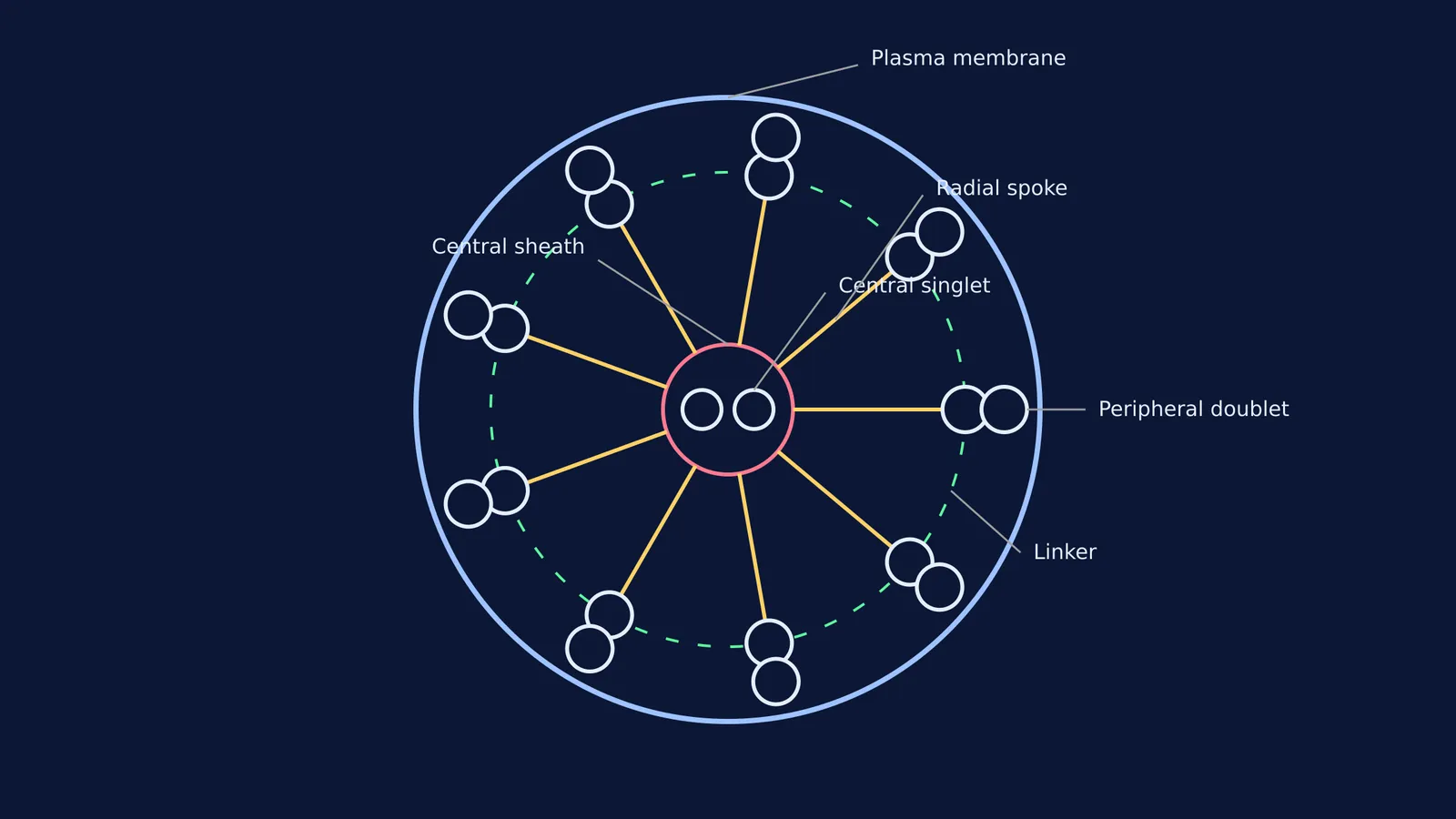
Their core, known as the axoneme, is supported by a highly organised bundle of microtubules. In this structure, there are nine pairs (doublets) of peripheral microtubules arranged in a ring, and two individual microtubules at the centre. This geometry is universally known as the 9+2 axoneme arrangement, as outlined in OpenStax Biology.
The central microtubules are enclosed by a sheath. Radial spokes bridge this central sheath to the nine peripheral doublets, while linkers connect the doublets to each other. This exact arrangement allows the microtubules to slide against one another, bending the cilium or flagellum and propelling the cell forward.
Why the cytoskeleton matters for NEET and board exams
For biology students, distinguishing the protein makeup and the structural arrangement of these three filaments is a frequent testing point. Questions in NEET and CBSE board exams often require students to match tubulin with microtubules, or actin with microfilaments.
Another common pitfall is confusing the microtubule geometry of different organelles. Remember that cilia and flagella exhibit a 9+2 arrangement, meaning nine peripheral doublets and two central singlets (OpenStax Biology). By contrast, a centriole displays a 9+0 arrangement made of nine peripheral triplets and no central microtubules (Wikipedia). Keeping these numbers straight is a simple way to secure marks in cell biology. If you find yourself repeatedly making mistakes on these factual distinctions, Fermi's AI tutor can work out what you already know and build a plan for your study session, checking your understanding as it goes.


