On this page
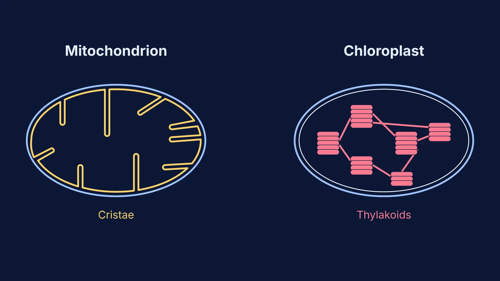
Mitochondria and chloroplasts are the powerhouses of the eukaryotic cell. While they handle different fuels—glucose for mitochondria, sunlight for chloroplasts—they share a striking structural feature: their inner membranes are highly folded. In mitochondria, these folds are called cristae, while in chloroplasts, they take the form of stacked, flattened sacs called thylakoids.
But why do these inner membranes fold in the first place, and what happens along their surfaces?
Why do inner membranes fold?
The primary reason for membrane folding is to increase surface area without increasing the overall volume of the organelle.
Energy production in both organelles depends on a series of membrane-bound proteins called the electron transport chain, as well as the enzyme ATP synthase. These proteins must be physically embedded in a membrane to function, because they pump protons (H⁺ ions) across it to build a gradient.
If the inner membrane were just a smooth sphere like the outer membrane, there would only be enough space for a small number of these protein complexes. By folding inwards repeatedly, the cell creates a massive functional surface packed with thousands of copies of the electron transport machinery.
What is the difference between cristae and thylakoids?
While both structures increase surface area, they belong to different organelles and have different layouts.
Mitochondria: Cristae
Mitochondria are bounded by two membranes. The outer membrane is smooth, while the inner membrane invaginates (folds inward) to form cristae. The space inside the inner membrane is called the matrix. The cristae house the proteins for oxidative phosphorylation—the final stage of aerobic respiration where most of the cell's ATP is made.
If you are revising cellular respiration, you can see how this fits into the broader picture in our guide on respiration as an amphibolic pathway.
Chloroplasts: Thylakoids
Chloroplasts also have a double-membrane envelope, but the folding happens in a separate, third membrane system inside. This thylakoid membrane is folded into disclike sacs that stack on top of each other like coins, forming structures called grana. The fluid surrounding the thylakoids is the stroma. The thylakoid membranes hold the photosystems (containing chlorophyll) that run the light-dependent reactions of photosynthesis.
How much does surface area increase?
The degree of folding directly reflects how much energy a cell needs.
According to *Molecular Biology of the Cell*, in a standard liver cell, the surface area of the mitochondrial inner membrane is about five times larger than that of the outer membrane. In cells with higher energy demands, such as cardiac (heart) muscle cells, the cristae are packed even more densely, creating an even larger surface area.
In chloroplasts, the scale is similarly impressive. The thylakoid membranes provide a vast expanse for capturing photons. (For more on how plants maximise their photosynthetic output, read about limiting factors in photosynthesis).
How do you remember the key features?
When comparing the two for exams, keep these distinctions clear:
Location: Cristae are in mitochondria; thylakoids are in chloroplasts.
Process: Cristae are the site of the electron transport chain in respiration; thylakoids are the site of the light-dependent reactions.
Internal Space: The space enclosed by cristae is the mitochondrial matrix; the space enclosed by thylakoid membranes is the thylakoid lumen.
If you need to review the individual steps of the electron transport chain or understand how the proton gradient forms, the Fermi Concept Library has every concept in biology, each with its own lesson and its place in the map.