

On this page
What is the difference between a common cold and pneumonia?
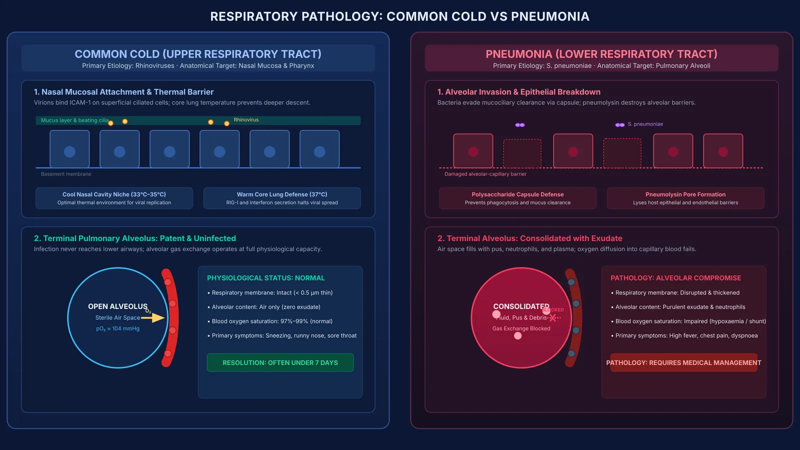
Although both conditions cause respiratory distress, their anatomical sites, pathogens, and physiological dangers differ fundamentally. The common cold is a self-limiting viral infection of the upper respiratory tract—chiefly the nose and throat. In contrast, pneumonia is an acute inflammatory infection of the lower respiratory tract, specifically the microscopic alveolar air sacs where blood exchanges oxygen and carbon dioxide.
According to the World Health Organization, pneumonia accounted for 14% of all deaths in children under five years old in 2019. Understanding this distinction is essential for biology students and NEET-UG aspirants. While viral particles irritating nasal mucosa cause transient discomfort, bacterial proliferation in terminal alveoli directly impairs arterial oxygenation.
Why does the common cold stay in the upper respiratory tract?
The restriction of the common cold to the upper airway stems from the thermal and molecular ecology of its primary pathogen. While multiple respiratory viruses can produce upper airway symptoms, rhinoviruses are the most frequent cause of the common cold, as documented by the Centers for Disease Control and Prevention.
Rhinoviruses replicate optimally in the cooler nasal passages, where inhaled air maintains mucosal temperatures between 33°C and 35°C. In a landmark study in the *Proceedings of the National Academy of Sciences*, Ellen F. Foxman and colleagues showed that airway cells mount a much stronger innate immune defense at lung temperature (37°C) than at 33°C. At core temperature, epithelial cells activate RIG-I-like receptor pathways that trigger rapid secretion of type I interferons, blocking rhinovirus replication before it reaches the lungs.
Because rhinoviruses bind primarily to intercellular adhesion molecule-1 (ICAM-1) on superficial nasal epithelial cells without invading deeper alveolar parenchyma, the lungs remain clear. The resulting symptoms—sneezing, rhinorrhea, nasal congestion, and mild sore throat—typically peak and often resolve in less than 7 days as mucosal defenses clear the viral infection. You can explore how these infectious agents fit into broader biological taxonomy in our guide to the classification of viruses and non-cellular agents.
How does bacterial pneumonia disrupt gas exchange in the alveoli?
Pneumonia presents an entirely different pathological scenario because the infectious agent reaches the fragile respiratory membrane of the terminal lung units. While viruses and fungi can cause pneumonia, bacterial species such as *Streptococcus pneumoniae* and *Haemophilus influenzae* represent the primary culprits in acute lobar pneumonia.
In a review in *The Lancet*, researchers Tom van der Poll and Steven M. Opal61114-4) described how *Streptococcus pneumoniae* evades upper airway clearance using its polysaccharide capsule. Reaching the alveoli, the bacteria release toxins like pneumolysin, damaging host membranes and triggering intense inflammation. Alveolar capillaries leak plasma, neutrophils, and red blood cells into the air sacs—a state known as alveolar consolidation.
In healthy lungs, oxygen diffuses across an ultrathin respiratory membrane into pulmonary capillaries. When inflammatory fluid fills the alveolar lumen, diffusion distance increases dramatically. As explained in our guide to breathing and alveolar gas exchange, this produces a severe ventilation-perfusion mismatch. Poorly oxygenated blood enters systemic circulation, causing dyspnoea, high fever, and, in severe cases, cyanosis—grayish-blue lips and nails caused by excessive deoxygenated haemoglobin. In laboratories, these bacterial pathogens are sorted using Hans Christian Gram's staining technique.
How can students analyze respiratory symptoms systematically?
When revising for exams or dissecting diagnostic biology problems, memorizing isolated symptoms often leads to confusion. Using a systematic physiological framework prevents common classification errors:
Identify the anatomical compartment: Determine whether the question describes the conducting zone (nasal passage, pharynx, trachea) or the respiratory zone (alveoli, alveolar ducts).
Characterize the pathogen's niche: Recall whether the organism thrives strictly in cooler mucosal passages (such as rhinovirus in the upper nasal epithelium) or invades deeper core tissues (*Streptococcus pneumoniae*).
Evaluate gas diffusion barriers: Check if the alveolar-capillary membrane is structurally preserved. In a cold, alveolar partial pressures of oxygen (pO₂) remain completely normal; in pneumonia, intra-alveolar exudate sharply lowers oxygen transfer into erythrocytes.
Interpret systemic markers: Nasal dripping and hoarseness point to superficial mucosal inflammation. Shaking chills, pleuritic chest pain, tachypnoea, and peripheral cyanosis point directly to alveolar consolidation and systemic hypoxaemia.
Consider a worked NEET comparison: a clinical question describes a patient presenting with high fever, rusty sputum, and dullness on chest percussion. Because fluid replaces alveolar air, sound vibrations transmit differently through consolidated tissue, while gas diffusion collapses. A student applying this framework immediately recognizes alveolar consolidation rather than upper airway rhinitis.
How Fermi supports active learning in biology
Mastering the connection between microscopic cellular mechanisms and macroscopic human pathology requires moving beyond passive textbook reading. The Fermi Concept Library organizes every concept across biology and physiology into an interconnected map, allowing you to trace how epithelial structures, membrane transport, and immune responses interact.
When practicing complex diagnostic questions and multi-step physiological problems, the Fermi Problem Library provides worked problems with a step-by-step solver that explains the reasoning rather than printing an answer. This structured approach helps students cultivate rigorous reasoning skills across high school and competitive examinations.


