
On this page
The cell is where biology starts, and the Class 8 version is mostly about seeing: preparing a slide, spotting the parts, and explaining why cells look different. This post covers the parts and their jobs, plant versus animal cells, the levels of organisation, one magnification calculation, and the slips that cost marks.
Where is the cell in the current book?
NCERT's Class 8 book is Curiosity, marked "Reprint 2026-27". Cells are in Chapter 2, "The Invisible Living World: Beyond Our Naked Eye", together with microorganisms. The older Class VIII book's 2024-25 reprint had no cell chapter at all, so if an older sibling's notes skip it, that is why. The topic continues in Class 9, which our guide to the cell in Class 9 covers.
Who first saw cells?
The chapter tells the story. In 1665 Robert Hooke published Micrographia. Looking at a thin slice of cork, he saw small empty compartments like a honeycomb and called each one a cell. Around the same time, in the 1660s, Antonie van Leeuwenhoek made better lenses and was the first to clearly see and describe tiny living things like bacteria and blood cells. Our post on Robert Hooke tells his part in more detail.
How do you see a cell in the lab?
Curiosity has two activities.
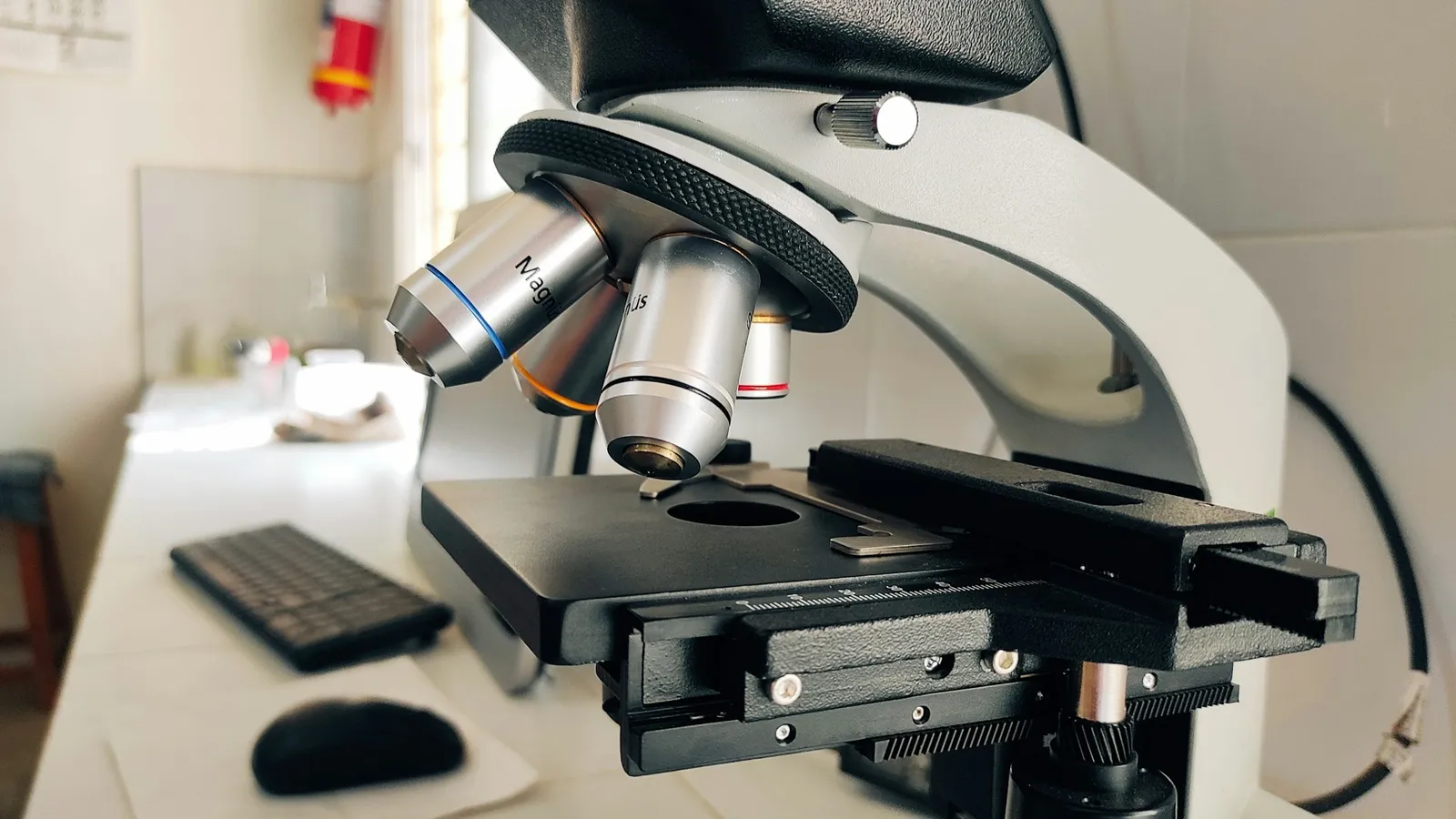
Onion peel: pull the thin layer from inside an onion scale, stain it with safranin for 30 seconds, rinse, mount it in a drop of glycerin and lower a coverslip without trapping air bubbles. You see nearly rectangular cells packed with no gaps, like bricks in a wall.
Cheek cells: gently scrape the inside of your cheek with the blunt end of a clean toothpick, spread it in water on a slide, add methylene blue, then glycerin and a coverslip. You see flat, irregular cells.
The stains add contrast so the parts show up. Glycerin stops the cells drying out.
What do the parts of a cell do?
The book names three basic parts found in all cells:
Cell membrane: the outer boundary. It separates one cell from another and is porous, letting in materials the cell needs and letting out waste.
Cytoplasm: fills the space between membrane and nucleus. Most life processes happen here.
Nucleus: a round structure with its own membrane. It controls the cell's activities and growth.
Plant cells add:
Cell wall outside the membrane, giving rigidity and strength.
Plastids, such as chloroplasts, which contain chlorophyll for photosynthesis.
A large vacuole, which stores substances and helps keep the cell's shape. Animal cells usually lack vacuoles, or have small ones.
Bacteria are different again: they have no well-defined nucleus, only a nucleoid region.
Why do cells have different shapes?
Shape matches job. Nerve cells are long and branched, so they can carry messages quickly to different parts of the body. Muscle cells are spindle-shaped and can contract and relax, which is how the food pipe pushes food down. Cheek cells are thin and flat and form a protective lining.
Cells then build up in levels: cell → tissue → organ → organ system → organism. A group of similar cells makes a tissue, tissues make an organ, and organs work together in a system.
Cell size varies hugely. The book notes that the yolk of an ostrich egg is a single cell, about 130 mm to 170 mm across.
Worked example: how big is the cell really?
Curiosity says a school microscope magnifies 100 to 400 times, and an electron microscope about 10,00,000 (one million) times.
Under the 400× setting, a cheek cell's image measures 20 mm across. What is its real size?
Real size = image size ÷ magnification = 20 mm ÷ 400 = 0.05 mm.
In micrometres (1 mm = 1,000 µm): 0.05 × 1,000 = 50 µm.
Now reverse it. A structure 0.0002 mm across, viewed at 10,00,000×, has an image of 0.0002 × 1,000,000 = 200 mm.
The usual mistake is to multiply when you should divide. A real cell must come out smaller than its image. If your answer is bigger, flip the operation.
Which mistakes cost marks?
Giving animal cells a cell wall. Only plant, fungal and bacterial cells have one.
Calling the cell membrane "solid" or sealed. The book calls it porous.
Mixing up the stains. Safranin for onion peel, methylene blue for cheek cells.
Skipping a level of organisation. Tissue comes between cell and organ.
A practice routine for this chapter
Day 1: Draw a plant cell and an animal cell from memory and label six parts in total.
Day 2: Write the job of each part in one line.
Day 4: Solve: an image 12 mm across at 400×. Find the real size (answer: 0.03 mm, or 30 µm).
Day 7: Explain to someone why a nerve cell is long and branched.
Self-testing like this tends to stick better than re-reading; a review of study techniques rated practice testing as high-utility and re-reading as low. If one part of the cell still feels vague, the Fermi Concept Library covers every concept in physics, chemistry, biology, mathematics and computing, so you can read that idea on its own.
Tonight, draw both cells from memory, then check your labels against Chapter 2.