

On this page
In biology, one of the most important rules is that form fits function. A cell's shape is never random; it is built specifically to do its job.
When you study Cell structure and functions in Class 8, you learn that while the smallest bacterial cells are just 0.1 to 5.0 micrometres across (Wikipedia)), cell shapes vary just as much as their sizes. From the long wires of your nervous system to the tiny discs in your blood, here is how four key cells match their shape to their work.
Why are red blood cells disc-shaped?
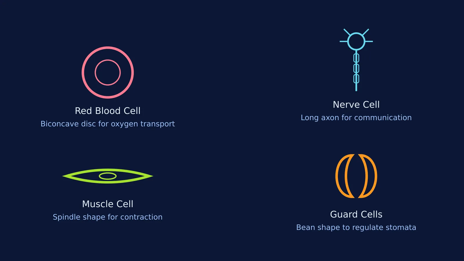
Red blood cells have a unique shape: they are biconcave discs, meaning they look like round cushions pinched in the middle.
This shape has two main benefits for transporting oxygen around the body (Wikipedia). First, it gives the cell a large surface area compared to its volume, allowing oxygen to diffuse in and out quickly. Hemoglobin, the protein that carries oxygen, fills the inside of the cell. The biconcave shape ensures that no hemoglobin molecule is far from the cell membrane, which cuts down the distance oxygen has to travel.
Second, the disc shape makes the cell highly flexible. A mature human red blood cell lacks a nucleus, freeing up even more space for hemoglobin and allowing the cell to squash into tight folds. This flexibility lets it squeeze through narrow capillaries that are even smaller than the cell itself, ensuring every tissue gets oxygen. You will look closer at their inner workings when you cover The cell in Class 9.
What is the function of a nerve cell's long branches?
A nerve cell, or neuron, looks like a tiny tree with a very long trunk. It has a central body with many short branches (dendrites) and one extremely long extension (the axon).
This shape is built for communication. The short branches let a single neuron receive chemical signals from thousands of its neighbours at once and convert them into electrical impulses. The long axon acts like an electrical wire to transmit these messages quickly over large distances.
To speed up the signal, the axon is often wrapped in a fatty layer called a myelin sheath, which acts like the rubber insulation on a copper wire. At the end of the axon, terminal branches pass the message to the next cell. This specialised wire-like shape allows your brain to tell your toe to move almost instantly. In fact, nerve cells are the longest cells in the human body; a single motor neuron reaching from your lower spinal cord to your big toe can be up to 1 metre long (Wikipedia).
How does a muscle cell's shape help it move?
Muscle cells are spindle-shaped or long and thread-like, meaning they are elongated and pointed at the ends.
This elongated shape is essential for their primary job: contraction (Wikipedia). A muscle works by shortening its cells to pull on bones or organs. Because muscle cells are arranged in long fibres, they have the physical length required to pull tightly together and relax again.
The long shape houses bundles of proteins called actin and myosin. During a movement, these protein filaments slide past one another, making the entire cell shorter and thicker. When the brain sends a signal to relax, the filaments slide back, and the spindle shape returns to its full length. Whether it is a skeletal muscle cell moving your arm, or a smooth muscle cell pushing food through your stomach, this thin structure is the only shape that allows for such dramatic changes in length. Without it, the muscle contraction step by step process of pulling and releasing could not happen.
Why are plant guard cells bean-shaped?
If you look at the surface of a leaf under a microscope, you will see tiny pores called stomata. Each stoma is surrounded by two guard cells, which in most plants look like two kidney beans facing each other.
Their job is to open and close the pore to let carbon dioxide in and water out (Wikipedia). Plants do not have a heart to pump fluids, so they rely on transpiration — the evaporation of water from leaves — to pull water up from the roots. Guard cells are the gatekeepers of this process.
The bean shape and their unevenly thickened walls make this mechanical response possible. Their inner walls (facing the stoma) are thicker and stiffer than their outer walls. When the plant has plenty of water, the guard cells swell. The thin outer wall expands more than the thick inner wall, forcing the cell into a curve and opening the gap. When the plant is dry, they shrink and straighten, closing the gap to save water.
If you find it hard to picture how these structures look in three dimensions, the Fermi Concept Library can help you map out the connections. You can explore how a single cell's shape fits into the larger tissue, and Fermi's AI tutor will explain the reasoning behind every biological design if you get stuck.


