

On this page
In multi-cellular organisms, cells do not just float next to each other in isolation. They are physically and chemically linked to form coherent tissues. These microscopic connections, known as intercellular junctions, act as structural anchors, waterproof barriers, and direct communication channels.
Understanding the difference between tight junctions, gap junctions, and adhering junctions is a core part of biology. It helps explain how tissues maintain their integrity and coordinate their functions, which is especially important if you are preparing for medical entrance exams.
What is the function of tight junctions?
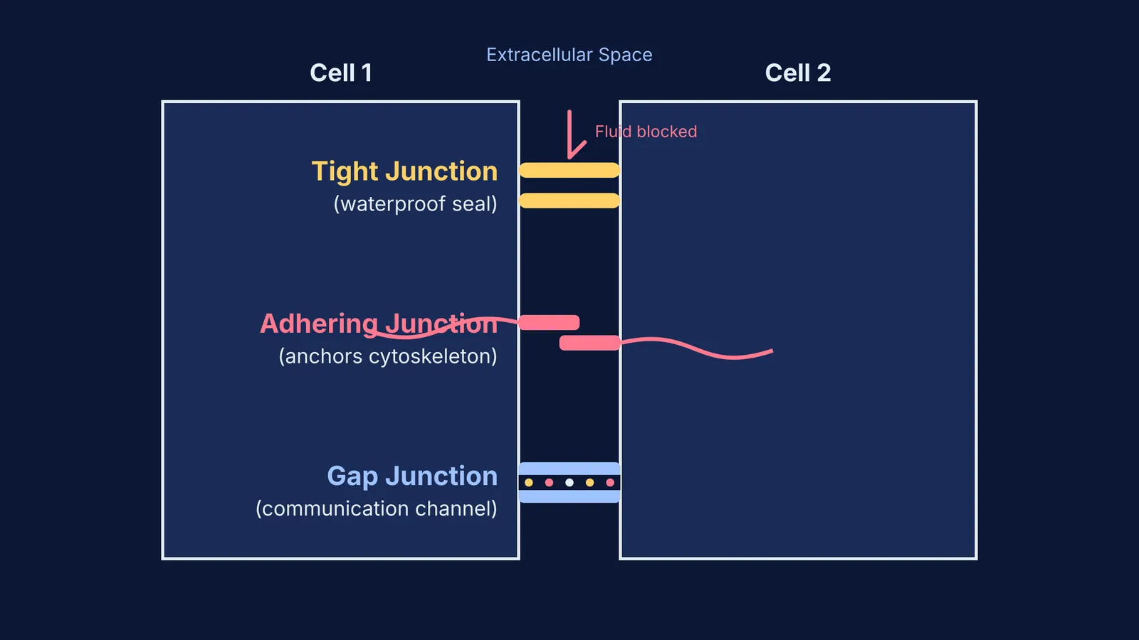
Tight junctions, or zonula occludens, are the body's waterproof seals. They fuse the plasma membranes of adjacent cells together, forming a continuous, tightly regulated barrier that prevents extracellular fluid from leaking through the microscopic spaces between cells.
This paracellular transport regulation is critical for maintaining an intact tissue barrier. In the digestive tract, tight junctions prevent intestinal contents and bacteria from leaking into the bloodstream. A failure of this regulation, often referred to as increased intestinal permeability, is linked to various immune and metabolic conditions (10.3389/fnut.2026.1938370). These junctions are primarily built from branching networks of transmembrane proteins, such as claudins and occludins, which stitch the two membranes together.
Beyond sealing, tight junctions also maintain the distinct "top" (apical) and "bottom" (basolateral) sides of a polarized epithelial cell. By acting as a fence, they stop membrane proteins from drifting between the two regions, ensuring that transport pumps face the correct direction.
What do adhering junctions do?
Adhering junctions, also known as adherens junctions, act as strong mechanical anchors. While tight junctions focus on sealing spaces, adhering junctions hold cells tightly together against severe physical stress and tearing.
These junctions couple intercellular adhesion molecules directly to the actin cytoskeleton inside the cell (10.1002/9780470015902.a0020876). The primary proteins involved are cadherins, which reach across the intercellular space and bind to identical cadherins on the neighboring cell in a calcium-dependent manner. Inside the cell, cadherins attach to the actin filaments via bridging proteins called catenins.
This creates a continuous transcellular network that provides immense structural integrity and is crucial for maintaining tissue shape. You will find them abundantly in tissues that must stretch, expand, and contract constantly, such as the epithelial layers of the skin and the endothelial lining of blood vessels. A related type of anchoring junction, the desmosome, functions similarly but connects to intermediate filaments like keratin instead of actin, functioning like spot welds to distribute shear forces.
How do gap junctions differ in function?
Unlike tight and adhering junctions, gap junctions are built purely for communication rather than physical strength or sealing. They form a functional syncytium by allowing neighboring cells to share their intracellular environments.
Gap junctions form tiny, hollow pores that bridge the narrow space between two cells. These channels allow ions, electrical signals, and small water-soluble molecules to pass directly from the cytoplasm of one cell into the next (10.1016/b978-0-12-802401-0.00011-9). Each pore is made of two half-channels, or connexons, contributed by the adjacent cells, which in turn are built from proteins called connexins.
This electrical and metabolic coupling is critical in tissues where rapid, coordinated responses are required. In the heart, gap junctions found within intercalated discs allow electrical action potentials to spread instantly across cardiac muscle cells, ensuring your heart beats in a coordinated, synchronized rhythm. They also play a major role in the central nervous system, acting as electrical synapses between neurons for near-instantaneous signal transmission.
How to organize your cell junction notes?
When revising Class 9 tissues or planning your NEET biology MCQ strategy, organize your study notes by function, defining protein, and primary location.
Identify the function: Ask yourself, "Does this specific tissue need to hold liquids, withstand mechanical stretching, or communicate rapidly?"
Match the protein: Link claudins and occludins to tight junctions, cadherins to adhering junctions, and connexins to gap junctions.
Use real-life locations: Picture the lining of the bladder (tight junctions preventing leaks), the epidermis of the skin (adhering junctions resisting abrasion), and cardiac muscle (gap junctions passing electrical signals).
Practice application: Test your recall using the Fermi Concept Library to see how these biological principles apply to complex, integrated physiology questions.
By understanding the distinct mechanical and chemical roles of these specialized junctions, the microscopic anatomy of human tissues makes logical sense rather than being a raw list of facts to memorize.


