

On this page
Every drop of soil water absorbed by a root must pass an anatomical checkpoint before reaching the xylem: the root endodermis and its gatekeeper, the Casparian strip. Discovered in 1865 by German botanist Robert Caspary, this band of hydrophobic polymer wraps around endodermal cells like a waterproof gasket. For students tackling school practicals or competitive exams, understanding where the strip sits and how it functions turns rote memorisation into clear, causal physiology.
Where does the Casparian strip sit in root anatomy?
The root is organized in concentric rings: epidermis on the outside, a broad cortex in the middle, and the central stele containing the vascular tissues. The endodermis forms the innermost boundary of the cortex, creating a single unbroken layer of barrel-shaped cells with zero intercellular spaces.
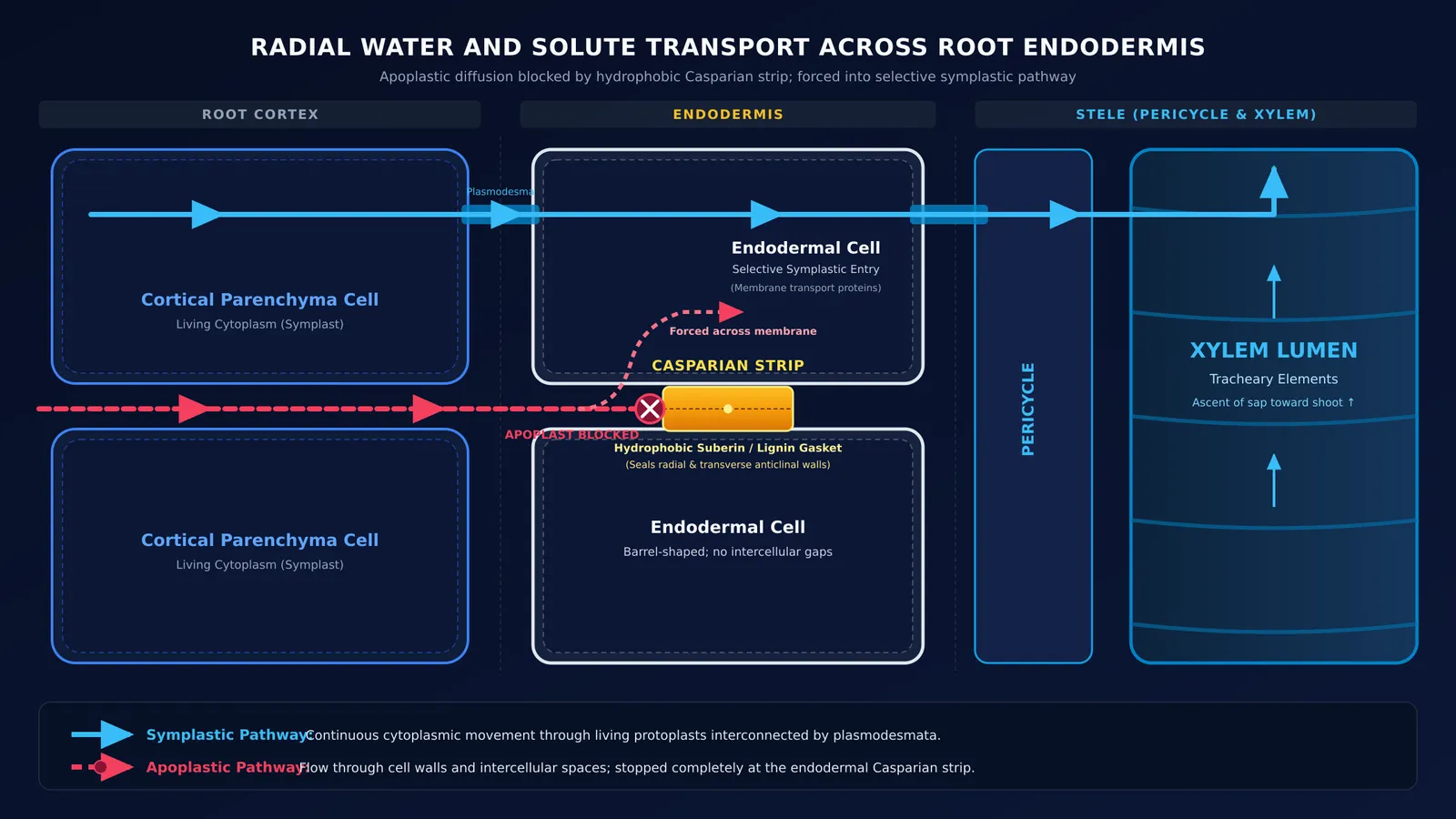
The Casparian strip does not coat the outer surface of these cells. Instead, it is embedded specifically within their anticlinal walls—the radial walls separating neighbouring endodermal cells and the transverse walls separating cells along the root axis.
Because the strip spans the primary wall and fuses tightly to the plasma membrane, it seals the gaps between adjacent cells. When reading diagrams in identifying plant anatomy transverse sections, look for this boundary line directly outside the pericycle. The Fermi Concept Library covers every concept across plant anatomy, each with its own lesson and its place in the map, showing how this boundary connects root structure to xylem transport.
What does suberin do to water and mineral transport?
Water moves through root tissue along two parallel pathways: the apoplast (through cell walls and intercellular spaces) and the symplast (through cytoplasm connected by plasmodesmata). The apoplastic route has low physical resistance, but it lacks selectivity. Without a barrier, unfiltered soil solutes and toxins would travel directly into the xylem stream.
The hydrophobic matrix of the Casparian strip prevents this by blocking apoplastic flow at the endodermis, as reviewed by Enstone et al. (2002). Water and solutes cannot squeeze through the cell walls; they are forced across the selectively permeable plasma membrane into the living symplast.
Once inside the cytoplasm, mineral uptake is regulated by transport proteins, ion channels, and aquaporins, echoing the principles of cell membrane transport mechanisms. The plant selectively admits vital ions like potassium and nitrate while restricting toxic elements. Furthermore, by preventing the back-leakage of xylem sap into the cortex, this seal maintains the osmotic gradient that powers root pressure.
Is the Casparian strip made of suberin or lignin?
Botanists long described the Casparian strip as a pure suberin band. However, research published by Naseer et al. (2012) in PNAS showed that the initial Casparian strip in Arabidopsis thaliana is made of lignin, not suberin. Plants engineered to lack suberin still formed functional strips that blocked apoplastic tracers, whereas blocking lignin synthesis dismantled the barrier. In a companion study, Roppolo et al. (2011) in Nature identified Casparian Strip Membrane Domain Proteins (CASPs) that direct localized lignin polymerization.
Suberin instead defines subsequent developmental stages:
Stage I: A localized Casparian strip made of lignin seals the apoplastic cleft.
Stage II: Suberin lamellae coat the inner face of the primary wall, sealing off transmembrane uptake in older root zones.
Stage III: Heavy U-shaped tertiary wall thickenings develop, as seen in monocot roots.
Even in Stage III, thin-walled passage cells remain opposite protoxylem poles to allow symplastic entry into the xylem.
How do you spot the endodermis and Casparian strip in a root section?
To identify the endodermis in a transverse section (T.S.) of a dicot or monocot root, use this four-step checklist:
Locate the cortex-stele boundary: Find the single, continuous ring of cells immediately enclosing the central vascular cylinder.
Check cell morphology: Endodermal cells are regular and barrel-shaped, packed edge-to-edge with no intercellular air spaces.
Find Casparian dots: Under a brightfield microscope, the cut radial walls appear as small, darkened, wedge-shaped thickenings known as Casparian dots.
Use diagnostic stains:
Phloroglucinol-HCl: Stains lignified Casparian dots and xylem vessels cherry red.
Berberine-aniline blue: As shown by Brundrett et al. (1988), berberine stains Casparian bands bright yellow-green under 365 nm ultraviolet light, while aniline blue quenches background fluorescence.
Fluorol Yellow 088: Fluoresces bright yellow under blue light specifically where suberin lamellae coat Stage II walls.
Recognizing these features helps resolve tricky assertions in NCERT-based biology questions. The Fermi Problem Library has worked problems with a step-by-step solver that explains the reasoning rather than printing an answer.


