

On this page
Why does your body need double circulation?
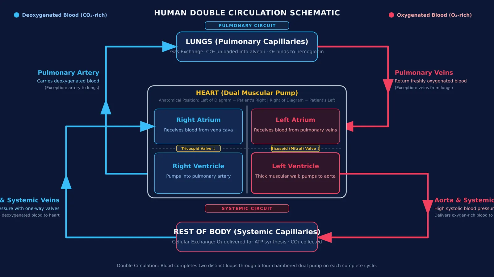
Every cell in your body needs a steady delivery of dissolved oxygen and glucose, along with an efficient way to clear carbon dioxide. In humans, this transport happens through double circulation, meaning blood travels through the heart twice during each complete bodily cycle. According to the NCERT Class 10 Science curriculum, warm-blooded animals require complete separation of oxygenated and deoxygenated blood to maintain constant body temperature and satisfy high metabolic energy demands.
As detailed in StatPearls cardiovascular research, the system operates through two linked circuits:
Pulmonary circulation: The right side of the heart sends deoxygenated blood to the lungs, where carbon dioxide diffuses out and fresh oxygen binds to hemoglobin.
Systemic circulation: The left side of the heart propels oxygenated blood through the aorta to tissues and organs across the body.
This closed-circuit concept was established in 1628 through William Harvey's proof that blood circulates in a continuous, one-directional loop driven by mechanical pumping.
How do arteries, veins, and capillaries compare?
Blood travels through three distinct types of blood vessels. As outlined in StatPearls vascular reviews, arteries and veins share three concentric wall layers: an inner endothelial lining (tunica intima), a middle smooth muscle layer (tunica media), and an outer connective coat (tunica adventitia).
Arteries: Arteries carry blood away from the heart under high systolic pressure. Their thick, muscular, elastic walls stretch and recoil with every ventricular beat.
Capillaries: Arterioles branch into microscopic capillary beds where nutrient exchange occurs. Capillary walls consist of a single endothelial cell layer with walls so thin and lumens so narrow that erythrocytes—explored in our guide to the formed elements of blood—must squeeze through in single file.
Veins: Veins collect deoxygenated blood from capillaries and return it to the heart under low pressure. Because venous pressure is low, veins feature wider lumens and one-way valves that prevent gravity from pulling blood backward.
Reviewing the contrast between elastic arteries and valved veins reinforces core questions in our Class 10 life processes transport guide.
How does blood move through the heart step by step?
The heart functions as a dual pump with four chambers: two thin-walled atria that receive blood and two thick-walled ventricles that pump blood outward. According to the StatPearls cardiac cycle reference, the left ventricle has the thickest muscular wall because it must generate enough force to supply the entire systemic circuit.
A single drop of blood follows an unvarying path:
Step 1: Deoxygenated blood enters the right atrium through the vena cava.
Step 2: Blood flows through the tricuspid valve into the right ventricle.
Step 3: The right ventricle pumps blood through the pulmonary artery to the lungs for gas exchange.
Step 4: Four pulmonary veins return freshly oxygenated blood to the left atrium.
Step 5: Blood passes through the bicuspid (mitral) valve into the left ventricle.
Step 6: The left ventricle contracts, ejecting blood into the aorta for systemic distribution.
How can you measure your pulse rate at home?
Every time your left ventricle contracts, it sends a pressure wave expanding the elastic walls of your arteries. You can palpate this pulse wherever an artery passes over a bone close to the skin surface.
According to the MedlinePlus Medical Encyclopedia, a normal resting heart rate for individuals aged 10 years and older ranges between 60 and 100 beats per minute. You can test your own pulse with this quick active-learning protocol:
Rest first: Sit quietly for at least 10 minutes so your cardiovascular system reaches baseline.
Locate the radial artery: Turn one hand palm up. Place the pads of your index and middle fingers on the wrist below the thumb base. Press gently until you feel a distinct beat. Avoid using your thumb, which has its own noticeable pulse.
Count the beats: Count the pulses for 30 seconds, then multiply by 2 to calculate your resting beats per minute.
Compare with exercise: Perform 60 seconds of jumping jacks or brisk step-ups, then recount your pulse immediately for 30 seconds and multiply by 2.
Guidelines from the American Heart Association explain that active muscles burn oxygen faster, prompting the autonomic nervous system to raise your heart rate to deliver more blood.
How do you master circulation diagrams for school exams?
Examiners frequently test whether students remember anatomical orientation. In diagrams, anatomical position is viewed facing the patient: the right atrium and right ventricle are drawn on the left side of the sheet, while the left chambers appear on the right.
Practice sketching the dual circuits from memory, using blue for deoxygenated pathways and red for oxygenated ones. The Fermi Concept Library maps every concept in biology to show how pulmonary gas exchange connects directly with cellular respiration. To watch pressure waves and valve mechanics operate dynamically, interactive models in the Fermi Simulation Lab help you visualize the flow patterns that static diagrams cannot convey.


