

On this page
The bacterial cell envelope is a multi-layered structure that determines the cell's shape and protects it from physical and chemical stress. Unlike viruses, viroids and lichens, all true bacteria possess a distinct and complex cellular envelope. If you are preparing for biology exams, understanding these layers is just as essential as knowing the structure of cilia and flagella because this architecture is the basis of the Gram stain and determines how antibiotics act.
Every bacterial cell envelope has a core set of layers, though their thickness and arrangement vary significantly between species.
What are the three layers of the cell envelope?
Moving from the outside of the cell towards the inside, the envelope typically consists of three distinct layers:
The Glycocalyx (Outer Layer)
This outermost "sugar coat" is composed of high-molecular-weight polysaccharides (Costerton et al., 1981). It primarily comes in two forms. The first is a well-organized, tightly bound capsule that acts as a major virulence factor, protecting the bacteria from being engulfed by immune cells (phagocytosis). The second is a looser, unorganized slime layer that easily washes off but helps bacteria stick to surfaces and form resilient biofilms.
The Cell Wall (Middle Layer)
The cell wall provides structural support to withstand the massive internal osmotic pressure of the cell. Its main ingredient is peptidoglycan (also called murein), a rigid, mesh-like polymer constructed of sugars and amino acids.
The Cell Membrane (Inner Layer)
Also known as the cytoplasmic membrane, this is a standard phospholipid bilayer. It acts as a selectively permeable barrier. Because bacteria lack internal organelles like mitochondria, this inner membrane also hosts the critical enzymes required for respiration, ATP synthesis and energy production.
If you struggle to picture how these structures stack together and function in three dimensions, you can explore the envelope model in the Fermi Concept Library.
Gram-positive vs. Gram-negative: What is the difference?
In 1884, Hans Christian Gram developed a staining technique that splits the bacterial world into two massive categories. We now know that this staining difference is not an arbitrary chemical reaction, but a direct result of the physical architecture of the cell envelope (Silhavy et al., 2010).
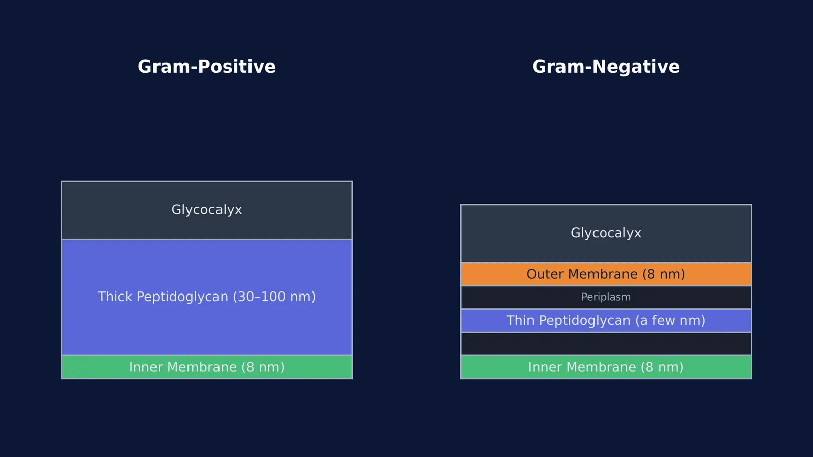
Gram-positive bacteria have a simple but highly robust envelope:
They possess a thick peptidoglycan layer, typically 30 to 100 nm deep (Silhavy et al., 2010).
Threaded heavily through this thick wall are teichoic acids (both lipoteichoic and wall teichoic acids), which give the cell surface an overall negative charge and help regulate cell division.
Crucially, they completely lack an outer membrane.
Gram-negative bacteria have a thinner, more complex "sandwich" structure:
They have a very thin peptidoglycan layer of just a few nanometers (Silhavy et al., 2010).
This thin wall is located in a gel-filled compartment called the periplasm, which sits between the inner membrane and a second, outer membrane.
The outer leaflet of this extra membrane contains lipopolysaccharides (LPS), a potent endotoxin. This outer membrane acts as a formidable physical barrier against large molecules, detergents and many antibiotics.
How does the Gram stain mechanism work?
The Gram stain specifically relies on the thickness of the peptidoglycan wall to either trap or release a dye complex during the washing phase (Tripathi and Sapra, 2025).
Here is the step-by-step mechanism of action:
Primary stain: Crystal violet, a cationic dye, is applied. It penetrates the cell envelope of both Gram-positive and Gram-negative bacteria, turning all cells purple.
Mordant: Iodine is added to the slide. The iodine binds with the crystal violet to form a large, insoluble complex (CV-I) that lodges within the peptidoglycan mesh.
Decolorization (The crucial step): An alcohol or acetone wash is applied. In Gram-positive cells, the alcohol severely dehydrates the thick peptidoglycan layer, causing its pores to shrink and permanently trap the massive CV-I complexes inside. In Gram-negative cells, the alcohol easily dissolves the lipid-rich outer membrane and damages the extremely thin peptidoglycan layer, washing the dye entirely out of the cell (Tripathi and Sapra, 2025).
Counterstain: Safranin is added to dye the now-colorless Gram-negative cells a contrasting pink or red.
A quick study checklist for bacterial layers
When revising this topic for your exams, make sure you can answer these four structural questions:
Is the capsule or slime layer part of the glycocalyx? Yes, both are forms of the glycocalyx polysaccharide coat.
Which type of bacteria has a thick peptidoglycan wall? Gram-positive bacteria have the thicker wall.
Where are lipopolysaccharides (LPS) found? They make up the outer leaflet of the outer membrane in Gram-negative bacteria.
What does the alcohol decolorizer do during a Gram stain? It dehydrates the Gram-positive wall to trap the dye, and dissolves the Gram-negative outer membrane to release it.
Understanding this architecture is the key to predicting bacterial behaviour, from how they survive in harsh environments to how doctors treat the infections they cause.


