

On this page
When you study the structure of a bacterial cell, you are looking at the defining example of a prokaryote. Unlike the eukaryotic cells that make up human skeletons or plants, bacteria lack a nucleus.
At 0.1 to 5.0 μm in diameter, prokaryotic cells are significantly smaller than eukaryotic cells (OpenStax). This tiny size is a structural advantage: a high surface area-to-volume ratio allows nutrients to diffuse quickly to all parts of the interior, and allows waste products to exit before they reach toxic levels. Because they are so small, bacteria rely heavily on their specialised structures to interact with their environment.
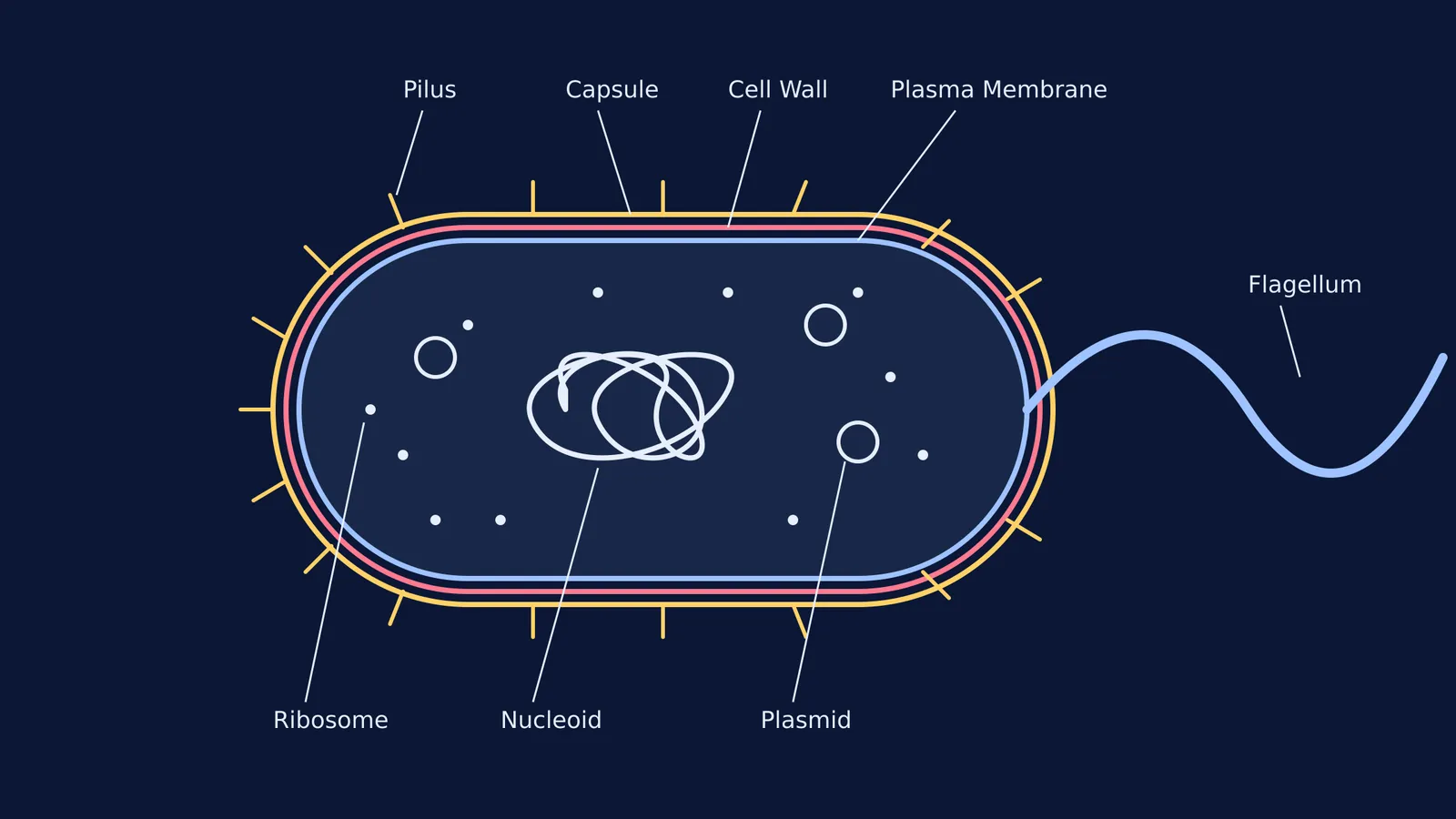
To draw and label a bacterial cell correctly for an exam, you need to place eight key structures accurately. Understanding what each part does will make it much easier to remember their positions on your diagram.
What are the three layers of a bacterial cell envelope?
The outside of a bacterium consists of up to three concentric layers, often called the cell envelope. From the outside in, they are:
Capsule: The outermost layer is a sticky, polysaccharide capsule. It enables the cell to attach to surfaces in its environment and offers protection from dehydration (OpenStax). In pathogenic bacteria, this layer also helps them evade the host's immune system.
Cell Wall: Just beneath the capsule lies the cell wall, which acts as an extra layer of protection and helps the cell maintain its shape. In bacteria, it is primarily made of a strong mesh-like material called peptidoglycan. This wall prevents the cell from bursting if it absorbs too much water. (Interestingly, many antibiotics, like penicillin, work by targeting this exact peptidoglycan layer).
Plasma Membrane: The innermost boundary is an outer covering that separates the cell's interior from its surrounding environment, controlling what enters and exits. It is a phospholipid bilayer, functioning similarly to the membrane in human cells.
Where is the DNA in a prokaryote?
Because prokaryotes lack membrane-bound organelles, their genetic material floats freely in the jelly-like cytoplasm. There are two places to label DNA:
Nucleoid: A prokaryote lacks a nucleus, so its main chromosomal DNA is located in a darkened central part of the cell called the nucleoid (OpenStax). Draw this as a single, large, tangled circular thread in the middle. It contains all the essential genes required for the bacterium's survival.
Plasmids: These are small, independent circles of DNA separate from the main chromosome. They often carry extra, non-essential genes, such as those for antibiotic resistance. They are highly mobile and can be shared with other bacteria.
If you are exploring how these genetic traits pass between organisms—or reviewing the differences between different algae classes and bacteria—the Fermi Concept Library maps out every concept in biology with dedicated lessons.
What are pili and flagella used for?
The appendages extending from the capsule serve specific active functions for the bacterium's mobility and communication:
Flagellum: Used for locomotion, a flagellum acts like a microscopic propeller (OpenStax). Draw it as a long, whip-like tail extending from one end of the cell. Some bacteria have one flagellum, while others may have multiple spread across their surface.
Pili: These hair-like structures are used to exchange genetic material during conjugation, the process by which one bacterium transfers genetic material to another through direct contact (OpenStax). Draw them as short bristles distributed uniformly around the capsule.
Fimbriae: Though less commonly asked for in basic diagrams, these are shorter bristle-like fibres used purely for attaching to surfaces, such as the lining of an animal's intestine.
Finally, the cell's interior is dotted with ribosomes. These structures synthesise proteins. While eukaryotic ribosomes are slightly larger, bacterial ribosomes perform the same crucial function: reading the genetic code and building the proteins the cell needs to function. In a diagram, scatter them as small dots throughout the cytoplasm, but ensure they do not cross the boundary of the plasma membrane.
How do you draw a bacterial cell diagram step by step?
When tackling a diagram in a biology exam, a methodical approach ensures you do not miss any marks. Follow this simple sequence:
Start with the envelope: Draw three large, concentric pill shapes. Label the innermost line "Plasma Membrane", the middle "Cell Wall", and the outermost "Capsule".
Add the appendages: Draw one long wavy line from one end for the "Flagellum". Add several short dashes all around the outer capsule and label them "Pili".
Fill the interior: Draw a densely tangled scribble in the centre to represent the "Nucleoid", and one or two small, neat circles nearby for the "Plasmids".
Complete with ribosomes: Add small dots floating in the empty space inside the inner membrane, labelling them "Ribosomes".
By practising this sequence, you can quickly recreate an accurate, fully labelled prokaryotic cell from memory.


