

On this page
What is the difference between autosomes and sex chromosomes?
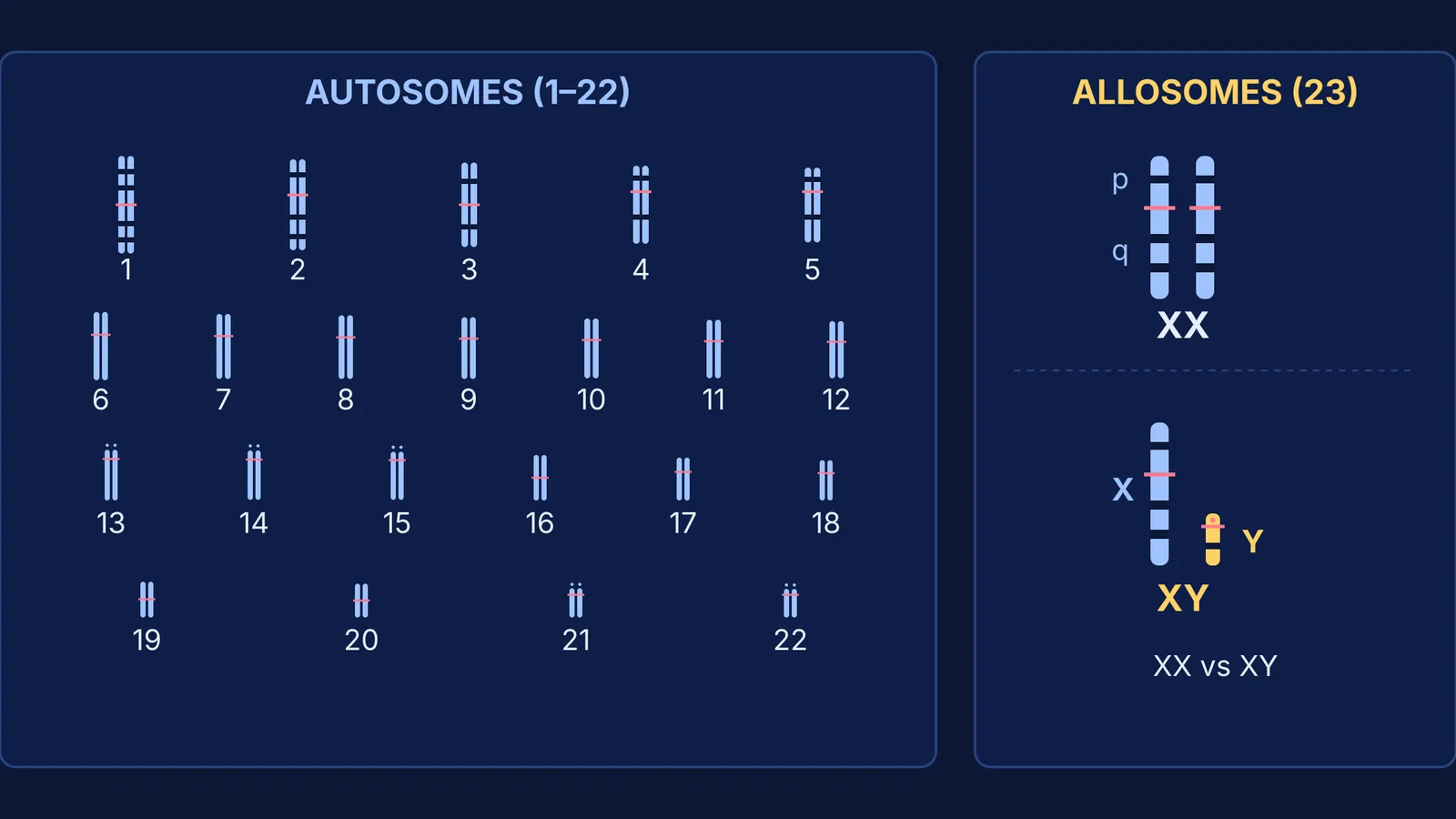
Every human nucleated somatic cell typically carries 46 chromosomes arranged in 23 pairs, a biological fact demonstrated by Joe Hin Tjio and Albert Levan in 1956 (Hereditas, DOI: 10.1111/j.1601-5223.1956.tb03010.x). These 23 pairs fall into two fundamentally distinct categories: autosomes and sex chromosomes (also called allosomes).
Autosomes comprise the first 22 pairs, or 44 chromosomes in total, according to the National Human Genome Research Institute Autosome Glossary. Autosomes are identical in general morphology across biological males and females. Each autosomal pair consists of true homologues: one chromosome inherited from the maternal parent and one from the paternal parent, carrying the same genes at identical loci in the same linear sequence. Autosomes carry genes responsible for standard somatic development, cellular metabolism, morphological structure, and enzymatic pathways.
In contrast, sex chromosomes constitute the single 23rd pair, or 2 chromosomes in total, as defined by the National Human Genome Research Institute Sex Chromosome Glossary. Sex chromosomes determine an individual's biological sex and carry sex-linked genetic traits. Unlike autosomes, sex chromosomes can be non-homologous: biological females typically inherit two X chromosomes (46,XX), forming a homologous pair, whereas biological males inherit one X chromosome and one Y chromosome (46,XY), forming a heteromorphic pair. The history of this realization traces back to early cytogenetic work, detailed in our account of Nettie Stevens's discovery of sex chromosomes.
How do gene counts and inheritance patterns compare?
The physical scale and genetic payload of autosomes and sex chromosomes differ markedly across the human genome:
Autosomes range broadly in physical size, roughly corresponding to their numeric designation. Chromosome 1 is the largest human chromosome, containing approximately 249 million base pairs and roughly 2,000 to 2,800 protein-coding genes according to the National Human Genome Research Institute. At the other end of the scale, chromosome 22 spans roughly 50.8 million base pairs, with Ian Dunham and colleagues reporting 545 genes in their landmark sequence (Nature, DOI: 10.1038/990031) and modern annotations confirming roughly 750 genes.
The X chromosome is large and gene-rich, spanning 155 million base pairs and containing roughly 800 to 1,000 protein-coding genes, sequenced by Mark Ross and colleagues in 2005 (Nature, DOI: 10.1038/nature03440). Genes on the X chromosome control diverse systemic traits that have nothing to do with sex determination, including blood clotting cascade proteins and retinal photopigments.
The Y chromosome is compact and gene-sparse, spanning roughly 57 million base pairs with a euchromatic region encoding around 60 to 70 protein-coding genes, sequenced by Helen Skaletsky and colleagues in 2003 (Nature, DOI: 10.1038/nature01722). Its master developmental switch is the SRY gene (sex-determining region Y), which triggers male testis differentiation during embryonic life.
Inheritance mechanisms follow these physical differences directly. Because autosomes exist as paired homologues in both sexes, autosomal genes follow standard Mendelian inheritance. Transmission ratios and phenotypic probabilities for autosomal traits are identical in sons and daughters, as seen in autosomal Mendelian disorders such as sickle-cell anaemia and PKU.
Sex-linked traits deviate from this symmetry. Because males possess only one X chromosome, they are hemizygous for X-borne loci. A single recessive mutant allele on the male X chromosome produces the mutant phenotype directly because no counterbalancing allele exists on the Y chromosome. In females, dosage compensation occurs via random X-chromosome inactivation, where one X chromosome condenses into an inactive Barr body during early development.
How does a laboratory prepare and read a human karyotype?
A karyotype is an organized laboratory display of an individual's condensed metaphase chromosomes arranged in homologous pairs. Cytogeneticists prepare karyotypes using living cells, most commonly peripheral blood lymphocytes, stimulated with phytohaemagglutinin to undergo cell division, as outlined by Nature Education Scitable.
The technical protocol relies on precise biochemical arrest and staining:
Cells in active division are exposed to colchicine or colcemid, which inhibits spindle fiber formation and traps cells at mitotic metaphase when chromosomes reach peak condensation.
The cell suspension is treated with a hypotonic potassium chloride solution, causing the cells to swell by osmosis and disperse their chromosomes across the glass slide upon dropping.
Slides are fixed with Carnoy's fixative (methanol and glacial acetic acid in a 3:1 ratio) and subjected to G-banding, a rapid trypsin-digestion and Giemsa staining method introduced by Marina Seabright in 1971 (The Lancet, DOI: 10.1016/s0140-6736(71)90287-x90287-x)).
G-banding produces an alternating pattern of light and dark bands along each chromatid. Dark bands reflect condensed, AT-rich heterochromatin with low gene density, whereas light bands indicate GC-rich, transcriptionally active euchromatin. Under a light microscope, digital software pairs homologues and organizes them into a standardized karyogram following the international Denver classification:
Group A (chromosomes 1 to 3): Large metacentric to submetacentric pairs.
Group B (chromosomes 4 and 5): Large submetacentric pairs.
Group C (chromosomes 6 to 12 and the X chromosome): Medium-sized submetacentric chromosomes.
Group D (chromosomes 13 to 15): Medium-sized acrocentric chromosomes bearing satellite stalks.
Group E (chromosomes 16 to 18): Short metacentric and submetacentric pairs.
Group F (chromosomes 19 and 20): Very short metacentric pairs.
Group G (chromosomes 21 and 22 and the Y chromosome): Small acrocentric chromosomes.
Reviewing chromosome types classified by centromere position helps students instantly distinguish these groups under high magnification.
How do you spot chromosomal differences in practice?
When interpreting karyotype plates in genetics examinations or laboratory assessments, following a systematic protocol avoids misidentifying normal variations as mutations:
Count total centromeres: Confirm whether 46 centromeres are present, referencing diagnostic guidelines from the National Human Genome Research Institute Karyotype Glossary. A count of 47 points to trisomy (such as Trisomy 21 in Down syndrome), while 45 indicates monosomy (such as Turner syndrome, 45,X).
Examine the 23rd position: Identify the sex chromosomes first. Two matching medium-sized submetacentric chromosomes confirm a 46,XX female karyotype. One medium submetacentric chromosome paired with a distinct, tiny acrocentric chromosome confirms a 46,XY male.
Compare autosomal pairs within Denver groups: Check that both homologues in pairs 1 through 22 share identical lengths, arm ratios (p arm to q arm), and matching G-band stripes.
Inspect for structural changes: Look for breaks, translocations between non-homologous autosomes, or segmental deletions along specific bands.
If you struggle to differentiate autosomal dominance from X-linked inheritance patterns on mock exams, the Fermi Problem Library offers worked problems with a step-by-step solver that explains the reasoning rather than printing an answer.
To master how centromeres, chromosome bands, and genetic loci fit into the broader human genome map, explore the Fermi Concept Library, which covers every concept in biology with its own lesson and its place in the map.


