

On this page
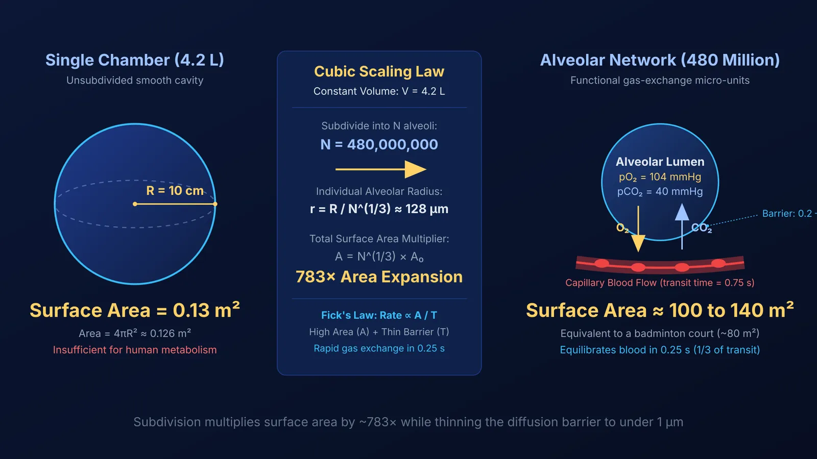
Every breath delivers oxygen to fuel trillions of cells. Human resting metabolism consumes roughly 250 mL of oxygen every minute, requiring continuous gas exchange between air and blood. If the adult lung volume of 4.2 litres were contained in two smooth, balloon-like chambers, their combined surface area would be only 0.13 square metres—scarcely enough to sustain a small reptile. Instead, design-based stereology led by Matthias Ochs in the *American Journal of Respiratory and Critical Care Medicine* (Ochs et al., 2004) shows that human lungs contain a mean of 480 million alveoli (ranging from 274 to 790 million), each averaging 4.2 × 10⁶ µm³ in volume.
Morphometric analysis by Peter Gehr, Marianne Bachofen, and Ewald R. Weibel in *Respiration Physiology* (Gehr et al., 197890104-4)) demonstrated that this subdivision creates an anatomical surface area of 143 ± 12 square metres, with an effective gas-exchange area of 70 to 100 square metres. As noted in Class 10 life processes, spreading out this alveolar surface covers approximately 80 square metres—matching the footprint of a regulation badminton court.
The geometry behind this gain follows cubic scaling:
A single sphere enclosing a lung volume of V = 4.2 × 10⁻³ m³ has radius R ≈ 0.10 metres (10 cm) and surface area 4πR² ≈ 0.126 square metres.
Dividing that volume into N = 480 million micro-spheres reduces individual radius to r = R / N^(1/3) ≈ 128 µm.
Total surface area equals N × 4πr² = N^(1/3) × 4πR², multiplying the area by 783 times to reach 98.7 square metres.
How does Fick's law govern alveolar diffusion?
Alveolar gas exchange relies entirely on passive physical diffusion. In 1855, Adolf Fick formulated the physical foundation of diffusion in *Annalen der Physik* (Fick, 1855), adapting Jean-Baptiste Joseph Fourier's heat conduction equations. Applied to pulmonary physiology, Fick's first law defines the rate of gas transfer (V_gas):
V_gas = (A / T) × D × (P₁ − P₂)
Here, A represents alveolar surface area, T is the thickness of the blood-gas barrier, D is the diffusion constant of the gas in the membrane, and (P₁ − P₂) is the partial pressure difference across the barrier. The diffusion constant depends on molecular solubility (S) and molecular weight (MW), governed by Graham's law of diffusion, scaling as D ∝ S / √MW.
In resting alveoli, oxygen partial pressure (pO₂) is roughly 104 mmHg, whereas deoxygenated blood arriving via the pulmonary artery has a pO₂ of 40 mmHg, creating a driving gradient of ΔP = 64 mmHg (OpenStax, 2022). By contrast, carbon dioxide partial pressure is 45 mmHg in arriving blood and 40 mmHg in alveolar air—a gradient of only 5 mmHg. Carbon dioxide diffuses just as rapidly despite this smaller gradient because its solubility in tissue fluids is 20 to 25 times higher than oxygen's, raising its diffusion constant.
What makes the respiratory membrane so thin and permeable?
A vast surface area would be ineffective if the barrier between air and blood were thick. Electron microscopy measurements by Gehr, Bachofen, and Weibel (Gehr et al., 197890104-4)) revealed that the harmonic mean thickness of the human tissue barrier is only 0.62 µm, with its thinnest segments measuring 0.2 to 0.3 µm. Because a red blood cell measures 7 to 8 µm across, the diffusion barrier is less than one-tenth the diameter of a single erythrocyte.
Three anatomical layers form this diffusion membrane:
The simple squamous epithelium of the alveolus, composed of thin Type I pneumocytes.
An acellular basement substance where epithelial and capillary basal laminae fuse.
The single-cell endothelial wall of the pulmonary capillary.
Centuries after Ibn al-Nafis and pulmonary circulation deduced that blood must flow through the lungs to reach the left ventricle, electron microscopy confirmed how closely air and blood meet.
To keep these micro-sacs from collapsing under surface tension, Type II pneumocytes secrete pulmonary surfactant. Laplace's law (P = 2γ / r) shows that smaller alveoli would generate higher collapsing pressures and empty into larger ones. Surfactant reduces surface tension in smaller alveoli, keeping all 480 million sacs stably inflated (Ochs et al., 2004).
Why is capillary blood transit time vital for oxygen saturation?
Alveolar architecture works because it is paired with a pulmonary capillary network spanning 126 square metres and holding 213 mL of blood (Gehr et al., 197890104-4)). In his quantitative analysis in *Respiration Physiology*, John B. West (West, 196990071-1)) showed that a red blood cell spends an average transit time of 0.75 seconds traversing a pulmonary capillary at rest.
Under resting conditions, oxygen equilibration between alveolar air and capillary blood is complete within 0.25 seconds—the first third of the erythrocyte's path (West, 196990071-1)). The remaining 0.50 seconds provides a three-fold diffusion reserve. During heavy exercise, when cardiac output rises and capillary transit time drops to 0.40 or 0.25 seconds, red blood cells still exit fully oxygenated.
Connecting physical formulas to biological structures is essential for mastering respiratory physiology. The Fermi Concept Library covers every concept in physics, chemistry, biology, mathematics and computing, each with its own lesson and its place in the map. For students tackling numericals on gas exchange, diffusion capacities, and partial pressures, the Fermi Problem Library provides worked problems with a step-by-step solver that explains the reasoning rather than printing an answer.


