

On this page
When a patient undergoes a PET scan to locate a tumour, the images are not generated by bouncing X-rays off their body. Instead, the patient is injected with a radioactive tracer that emits radiation from the inside out. Handling these tracers, operating the scanners, and ensuring everyone's safety is the job of a nuclear medicine technologist.
If you are drawn to a medical career that relies heavily on applied physics and chemistry, the nuclear medicine technologist course after 12th offers a direct route into this highly specialised field.
What is the nuclear medicine technologist course after 12th?
To become a technologist in India, you must complete a bachelor's degree, usually titled B.Sc. in Nuclear Medicine Technology. The programme typically includes academic study followed by a mandatory clinical internship.
During the course, students bridge the gap between biology and physics. You must have passed your 12th-grade board exams with Physics, Chemistry, and Biology (some institutes also accept Mathematics). The curriculum covers human anatomy, radiation physics, radiochemistry, and the operational mechanics of complex machines like PET (Positron Emission Tomography) and SPECT scanners.
If you find yourself struggling to visualise the atomic physics required for these entrance exams, Fermi's AI tutor can work out what you already know and build a plan for the session, teaching you the foundational decay equations step by step.
How do radioactive tracers work?
The magic of nuclear medicine lies in radiopharmaceuticals. One of the most common tracers is Fluorodeoxyglucose (FDG), a glucose analogue tagged with the radioactive isotope Fluorine-18. Because cancer cells consume glucose rapidly, the tracer accumulates in tumours.
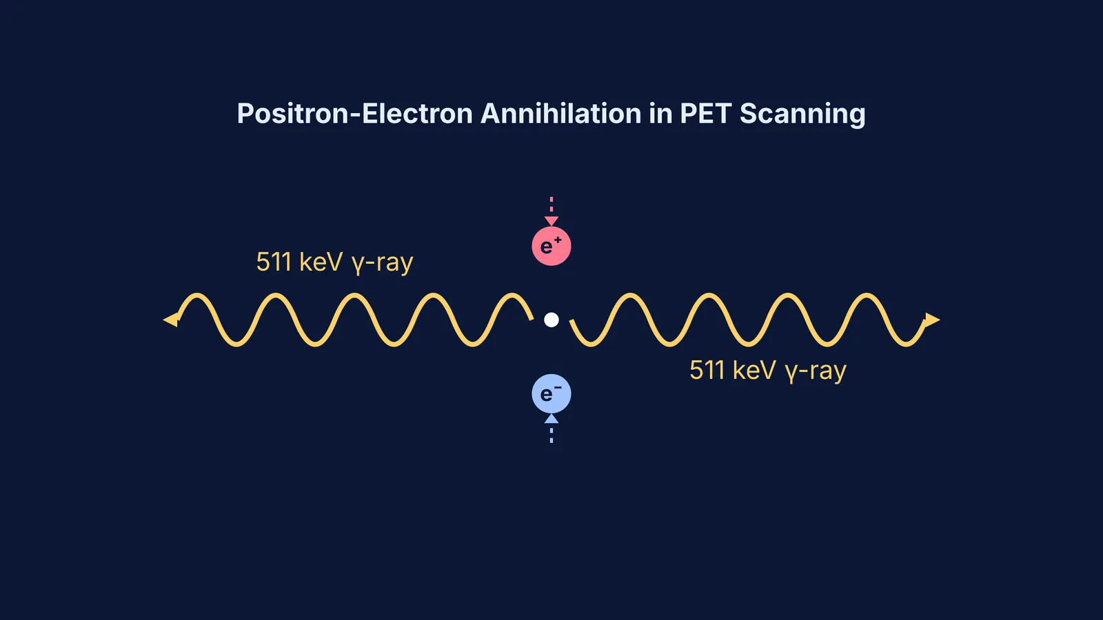
When the Fluorine-18 nucleus undergoes beta-plus decay, it emits a positron. This positron travels a very short distance before colliding with an electron in the patient's tissue. The two particles annihilate each other, converting their mass entirely into energy and emitting two 511 keV gamma rays in exactly opposite directions. The scanner ring detects these simultaneous gamma rays to pinpoint the location of the annihilation.
George de Hevesy and radioactive tracers in living things laid the historical groundwork for this technique, proving that radioactive isotopes could track biological processes without disturbing them. You can also explore how parent isotopes break down over time when counting alpha and beta decays in a radioactive decay series.
The challenge for the technologist is time. The physical half-life of Fluorine-18 is approximately 109.8 minutes). This means the tracer loses half its radioactivity in under two hours. Technologists must calculate the exact dose, verify the timing, and image the patient efficiently before the signal fades. To truly understand how this rapid decay changes the activity level minute by minute, you can run virtual decay experiments in the Fermi Simulation Lab.
What is the ALARA principle?
Because technologists work with unsealed radioactive sources daily, radiation safety is the most critical part of the training. In India, the training standards and facility operations are heavily regulated by the Atomic Energy Regulatory Board (AERB).
Technologists are trained to follow the ALARA principle—keeping radiation doses "As Low As Reasonably Achievable".
Time: Minimise the time spent near the radioactive source or the injected patient.
Distance: Maximise the distance from the source. Because radiation intensity drops with distance, stepping further back significantly reduces your exposure.
Shielding: Use dense materials to absorb the radiation. Technologists use tungsten or lead syringe shields when drawing up doses, and stand behind lead-glass barriers during the scan.
What is a technologist's daily workflow?
If you are considering this path, here is a practical checklist of what a technologist's routine looks like:
Morning calibration: Run quality control tests on the PET or SPECT scanners using standard radioactive sources to ensure the detectors are precise.
Dose preparation: Receive the radiopharmaceutical from the cyclotron facility, calculate the patient-specific dose based on their weight, and draw it up behind lead shielding.
Patient preparation: Explain the procedure to the patient, inject the tracer, and monitor them during the uptake time while the tracer circulates.
Image acquisition: Position the patient perfectly inside the scanner and operate the console to acquire the images.
Waste management: Store used syringes and gloves in heavily shielded bins until their radioactivity decays to background levels before disposal.
The nuclear medicine technologist course after 12th is not just another degree; it is a commitment to precision, patient care, and the invisible laws of physics.


