

On this page
Memorizing cross-sections of plant tissues can feel like staring at abstract art. However, knowing the exact structural rules is a core requirement for botany and medical entrance exams. When you ask AI dicot vs monocot root stem anatomy differences, you can dynamically test your knowledge instead of passively staring at a textbook. This approach forces you to recall the physical arrangement of tissues—like the xylem, phloem, and pith—before you look at the answer.
What are the main dicot vs monocot root differences?
The root system absorbs water and anchors the plant, but the internal plumbing differs drastically between plant groups. A dicot root typically has 2 to 8 vascular bundles, whereas a monocot root has 8 to many (BYJU'S). This higher number of bundles in monocots is known as a polyarch condition.
Another defining feature is the central core, or the pith. In a monocot root, the pith is large and well-developed (BYJU'S). By contrast, a dicot root has an inconspicuous or entirely absent pith. Furthermore, dicot roots undergo secondary growth. Their pericycle gives rise to the vascular cambium and cork cambium, allowing the plant to increase in girth over time. Monocot roots, on the other hand, do not exhibit secondary growth, which is why monocot plants like grasses or palms maintain a relatively uniform root thickness.
What are the differences between dicot and monocot stem anatomy?
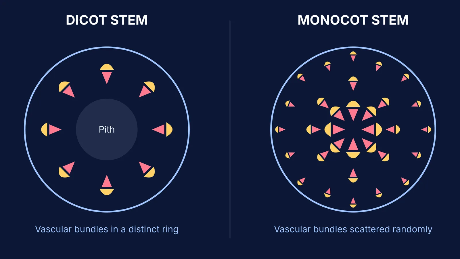
When you examine a cross-section of a stem under a microscope, the arrangement of vascular tissues is the most immediate giveaway. In a dicot stem, the vascular bundles are arranged in a distinct ring (OpenStax). This organized ring structure allows for the presence of a cambium between the xylem and phloem. Because of this cambium, dicot stems are described as having open vascular bundles, enabling the secondary growth that creates woody trunks in trees.
Conversely, a monocot stem has its vascular bundles scattered randomly throughout the ground tissue (OpenStax). These bundles are typically larger near the center and smaller towards the periphery. Because these scattered bundles lack a cambium, they are closed—meaning monocot stems do not undergo secondary growth. This scattered arrangement also means that the ground tissue in a monocot stem is an undifferentiated mass, whereas a dicot stem is clearly divided into distinct layers: the cortex, endodermis, pericycle, and pith.
How to check plant anatomy details against NCERT diagrams?
Standard textbook figures, like those found in the NCERT syllabus, pack a lot of overlapping labels into a small space. It is easy to look at the colorful diagram of an epidermis and endodermis and assume you have memorized it. But recognizing a diagram is not the same as recalling it from scratch. An interactive session with an AI can expose these gaps in your visual memory.
If you struggle to mentally map the layers from the outer hypodermis to the inner stele, Fermi's AI tutor can work out what you already know and build a plan for the session. It teaches rather than simply printing out a list of differences. It draws the figure on its board step by step, asking you to predict where the endodermis or pericycle should go before it completes the sketch.
If you need to review the definitions of these specific tissues before testing yourself—for instance, remembering whether the hypodermis is collenchymatous or sclerenchymatous—you can look them up in the Fermi Concept Library.
A practical framework for checking your knowledge
To get the most out of a study session, do not just ask the AI to summarize the chapter for you. Use this structured approach to simulate a practical viva exam:
Hide the textbook diagram and open your AI prompt.
Specify the exact cross-section you are testing (for example, a monocot root or a dicot stem).
Instruct the AI to move directionally from the outside in (epidermis to pith), requesting your input at each individual step.
Make sure to define the curriculum standard, such as NCERT, so the terminology matches your exam perfectly.
By explicitly asking the AI to verify these layers interactively, you transform static memorization into an engaging active recall exercise.


