On this page
Spirogram questions in NEET Biology can look intimidating when presented as oscillating curves. You are asked to pinpoint tidal volume, calculate vital capacity, or predict what shifts when a runner sprints. But if you understand what each wave represents on the instrument drum, reading a spirometer trace becomes intuitive.
What does a spirometer trace actually record?
A spirometer measures the volume of air moved into and out of your lungs. In 1846, English surgeon John Hutchinson's clinical study introduced the water-sealed spirometer to measure what he termed "vital capacity." Modern testing guidelines from the American Thoracic Society and European Respiratory Society (ATS/ERS 2019 standards) continue to rely on these tracings to assess pulmonary mechanics.
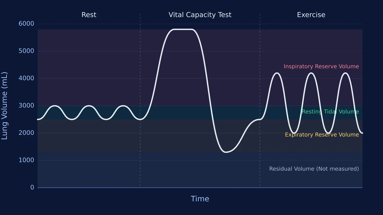
On a standard spirogram, time runs horizontally and lung volume runs vertically. An upward deflection marks inhalation as air enters the lungs, while a downward slope marks exhalation. The baseline oscillations represent quiet, relaxed breathing, bordered by reserve volumes your thoracic muscles recruit during exertion.
How do you extract lung volumes from a spirogram?
According to the NCERT Class 11 Biology curriculum (Chapter 17, Breathing and Exchange of Gases), a normal spirogram reveals four primary dynamic volumes:
Tidal Volume (TV): The amplitude of normal quiet breathing, averaging roughly 500 mL in a healthy adult (delivering 6000 to 8000 mL of air per minute).
Inspiratory Reserve Volume (IRV): The extra air you can inhale above normal end-inspiration, averaging 2500 to 3000 mL.
Expiratory Reserve Volume (ERV): The additional air you can forcefully expel below normal end-expiration, averaging 1000 to 1100 mL.
Vital Capacity (VC): The maximum air expelled after deepest inspiration (VC = ERV + TV + IRV), spanning 3500 to 4500 mL.
To review step-by-step calculations and gas exchange numericals, see our breathing and gas exchange revision guide.
Which lung volumes change when you exercise?
During exercise, your ventilation climbs significantly as you breathe faster and deeper. But vital capacity does not change.
Vital capacity is an anatomical ceiling determined by chest wall compliance and lung size. As Henke and colleagues showed in the Journal of Applied Physiology, active expiration during heavy exercise lowers end-expiratory lung volume by an average of 0.71 L, optimizing diaphragmatic recoil.
To increase ventilation, tidal volume expands by encroaching into both the inspiratory reserve volume and the expiratory reserve volume. Because tidal volume expands into both reserves, IRV and ERV shrink. Meanwhile, total lung capacity and vital capacity remain constant.
Why is residual volume invisible on a spirometer?
Even during maximal exhalation, your lungs do not collapse. According to the NCERT Class 11 Biology curriculum, around 1100 to 1200 mL of air remains trapped inside non-collapsible alveoli and airways. This is your Residual Volume (RV).
Because residual air never exits the chest, a spirometer drum cannot record it. Consequently, capacities that include residual volume—Functional Residual Capacity (FRC = ERV + RV) and Total Lung Capacity (TLC = VC + RV)—cannot be measured using standard spirometry alone. They require helium dilution or body plethysmography. Practising drawing NCERT biology diagrams from memory helps lock in which capacities include RV.
How do you predict reserves and exercise shifts?
Suppose an exam problem gives a spirogram with these volume levels:
Quiet inspiration peak: 3000 mL
Quiet expiration trough: 2500 mL
Maximal forced inspiration peak: 5800 mL
Maximal forced expiration trough: 1300 mL
From these levels:
TV: 3000 mL − 2500 mL = 500 mL
IRV: 5800 mL − 3000 mL = 2800 mL
ERV: 2500 mL − 1300 mL = 1200 mL
VC: 5800 mL − 1300 mL = 4500 mL
Now imagine the subject begins running: tidal volume expands to 2200 mL, and active expiration lowers the end-expiratory level to 2000 mL. What are the new reserves?
New ERV: 2000 mL − 1300 mL = 700 mL (reduced from 1200 mL).
New End-Inspiratory Level: 2000 mL + 2200 mL = 4200 mL.
New IRV: 5800 mL − 4200 mL = 1600 mL (reduced from 2800 mL).
Verification: TV (2200) + ERV (700) + IRV (1600) = 4500 mL. Vital capacity remains completely unchanged.
Educational research by Carl Wieman and colleagues in Science demonstrates that manipulating dynamic models helps students master physiological relationships far better than memorising static figures. You can also read more about Carl Wieman's research on active learning.
To see how changing airway resistance and exercise intensity shift these waveforms interactively, explore the Fermi Simulation Lab, which provides interactive simulations for the ideas that are easier to see than to read. You can review respiratory anatomy across the Concept Library, and solve multi-part capacity questions in the Fermi Problem Library, with a step-by-step solver that explains the reasoning.