

On this page
Spinach leaves look uniformly green, but that color hides a mixture of several chemical compounds. By using paper chromatography, you can split this mixture apart to see the distinct pigments that drive photosynthesis.
What is leaf pigment chromatography?
Chromatography separates a mixture by passing it through a medium where its components move at different rates. In paper chromatography, the extract is spotted onto a strip of filter paper (the stationary phase) and placed in a liquid solvent (the mobile phase).
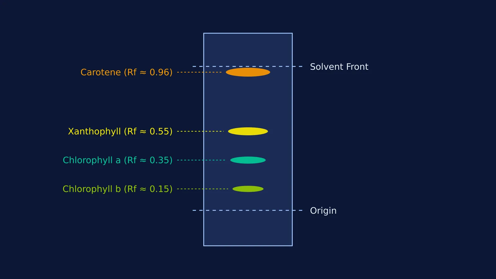
According to the official NCERT Class 11 Biology curriculum, a spinach extract separated this way reveals four specific bands from top to bottom:
Carotenoids (Carotene): Yellow to yellow-orange
Xanthophylls: Yellow
Chlorophyll a: Bright or blue-green
Chlorophyll b: Yellow-green
This separation works on the principle of polarity. The solvent carries the pigments up the paper, but each pigment clings to the paper with different strength. Water is highly polar, which is why it is not used as a mobile phase in leaf pigment chromatography; it would bind too strongly to the cellulose.
Why are there multiple pigments in a spinach leaf?
Chlorophyll a is the primary engine of photosynthesis, converting light energy into chemical energy. However, it only absorbs strongly in the red and blue regions of the visible spectrum. If a plant only had chlorophyll a, it would waste a large portion of the sunlight hitting its leaves.
This is where accessory pigments come in. Chlorophyll b, xanthophylls, and carotenes absorb light at different wavelengths—mostly in the blue and green regions. They capture this incoming energy and funnel it down to the chlorophyll a molecules at the reaction center.
By spreading the absorption range across the spectrum, these accessory pigments ensure that the plant maximizes energy capture even during the early morning or under dense forest canopies. When plants operate in different light conditions or face stress, they rely heavily on these accessory pigments to protect their photosynthetic machinery. For instance, understanding pigment roles is useful when studying how plants manage energy during Photorespiration: why RuBisCO wastes energy in C3 plants.
How do you calculate Rf values for spinach pigments?
To quantify the separation, scientists calculate a retardation factor (Rf) for each pigment.
The Rf value is the distance traveled by the pigment divided by the distance traveled by the solvent front. Both measurements start from the origin line where you first spotted the extract.
In a standard solvent system like petroleum ether and acetone, the pigments reliably separate in a predictable order. A study published in the Journal of Chemical Education (Quach et al., 2004) confirms that highly non-polar pigments travel furthest in mostly non-polar solvent mixtures.
Because conditions like humidity, solvent ratios, and paper type vary, the exact numerical Rf values will shift from lab to lab, but their relative order remains constant on paper: carotene travels near the solvent front (Rf > 0.90), followed by xanthophylls, chlorophyll a, and chlorophyll b.
Why does chlorophyll b travel the shortest distance?
The differences in Rf values come down to the molecular structure of the pigments. Chromatography paper is made of cellulose, which is highly polar. The solvent is mostly non-polar.
Carotene is a pure hydrocarbon without any oxygen atoms. Because it is highly non-polar, it ignores the polar paper and rides the non-polar solvent almost to the very top.
Chlorophyll b, by contrast, contains a highly polar aldehyde group. This makes it bind tightly to the polar cellulose fibers in the paper, resisting the upward pull of the solvent. Chlorophyll a has a less polar methyl group instead of an aldehyde, so it travels slightly further than chlorophyll b.
If you want to review the organic chemistry behind these functional groups and how they dictate solubility, the Fermi Concept Library maps out the molecular structures step by step.
Leaf pigment chromatography virtual lab: A step-by-step framework
Whether you are in a physical classroom or running a virtual lab, a structured approach prevents common errors like washing away the spot. Here is a checklist for running the experiment:
Extract the pigments: Grind the spinach leaves thoroughly in a mortar with a few drops of acetone to break down the cell walls.
Spot the origin: Draw a pencil line 2 cm from the bottom of the paper. Use a capillary tube to place a tiny, concentrated spot of extract on the line. Let it dry completely before applying more.
Run the chromatogram: Lower the paper into the solvent chamber, ensuring the solvent level is below the pencil line. Seal the chamber to saturate the air with vapor.
Mark the front: Remove the paper just before the solvent reaches the top edge. Immediately mark the solvent front with a pencil before it evaporates.
Measure quickly: Trace the outline of the pigment bands, as colors like xanthophyll will fade when exposed to air and light.
Visualizing capillary action and solvent dynamics can be difficult from a textbook alone. The Fermi Simulation Lab provides interactive simulations for the ideas that are easier to see than to read.


