On this page
The discovery of the DNA structure is one of the most famous stories in biology. It is often taught as a stroke of genius, but it was actually a masterclass in synthesis. James Watson and Francis Crick did not conduct the experiments that proved DNA's shape. Instead, they built a physical model that satisfied every constraint discovered by other researchers.
Why did Watson and Crick build a physical model?
In the early 1950s, scientists knew that DNA carried genetic information, but its three-dimensional shape was a mystery. Researchers like Linus Pauling were trying to solve it using physical models—literally building structures with wire and metal plates to see how atoms could fit together without breaking chemical laws.
Watson and Crick adopted this approach. They knew that a successful model had to account for two major pieces of independent evidence: the chemical composition rules found by Erwin Chargaff and the X-ray diffraction images produced by Rosalind Franklin.
How did Chargaff's rules point to base pairs?
In 1950, biochemist Erwin Chargaff published a paper analysing the chemical makeup of DNA. He found that while the overall sequence of bases varies between species, the ratios of the bases follow strict rules. Specifically, the number of adenine (A) units always equals the number of thymine (T) units, and the number of guanine (G) units equals the number of cytosine (C) units (10.1007/bf02173653).
For Watson and Crick, Chargaff's rules (A=T and G=C) were the key to the molecule's interior. They realised that if an adenine on one strand always bonded with a thymine on the other, and guanine always bonded with cytosine, the two strands would hold together perfectly.
If you want to practice the mathematics of these base pairs, try our guide on Chargaff's rule numericals. To understand the building blocks themselves, read up on nucleosides vs nucleotides.
What did Rosalind Franklin's Photo 51 reveal?
While Chargaff provided the chemical rules, Rosalind Franklin provided the physical dimensions. Working at King's College London in 1952, Franklin and her student Raymond Gosling produced "Photo 51", an exceptionally clear X-ray diffraction image of DNA.
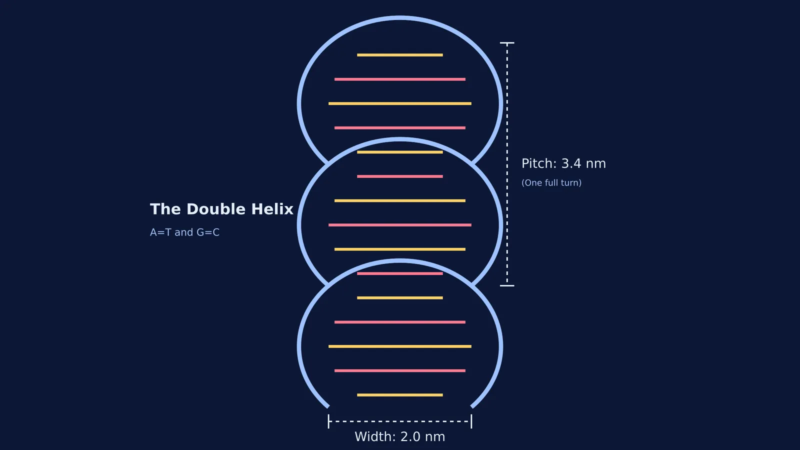
When Watson saw the image, the distinctive "X" pattern immediately told him the molecule was a helix. More importantly, Franklin's data provided the exact measurements needed to build the model: a width of 2.0 nanometres and a repeating pitch of 3.4 nanometres per turn (PMID: 13054694).
The 1953 model: what did they get right?
Before they got it right, Watson and Crick built several failed models. At one point, Watson tried pairing adenine with adenine and cytosine with cytosine, but the sizes did not fit the uniform width that Franklin's data demanded. Furthermore, Watson initially used the wrong chemical structures (the enol forms) for the bases.
It was only when a fellow chemist, Jerry Donohue, pointed out that the bases exist in a different shape (the keto form) under biological conditions that the puzzle pieces finally clicked. In their keto forms, adenine and thymine formed perfect hydrogen bonds, as did guanine and cytosine.
On 25 April 1953, Watson and Crick published their landmark paper in *Nature* (NLM ID: 101584582X381). Their model correctly placed the sugar-phosphate backbones on the outside, running in opposite directions (antiparallel), with the complementary base pairs meeting in the middle like the rungs of a twisted ladder.
Their synthesis answered both the structural and the functional questions of biology. As they famously noted at the end of their paper, the specific base pairing immediately suggested a mechanism for how DNA could be copied. To see where the search for this molecule began, you can read about Friedrich Miescher's discovery of nuclein.
A framework for model-building in your own study
Watson and Crick solved DNA by gathering constraints and building a model to test them. You can use a similar approach when tackling hard science problems:
List your constraints: Before you try to solve a problem, write down everything the final answer must satisfy. In physics, this might be the conservation of energy; in biology, it might be the base-pairing rules.
Build a mental or physical model: Draw the forces, map out the reaction, or sketch the cell structure. When you are trying to picture complex 3D structures or dynamic processes, reading text is often not enough. This is where the Fermi Simulation Lab helps, offering interactive simulations for the ideas that are easier to see than to read.
Check the details: Donohue's correction of the base structures reminds us that small details matter. Always verify the assumptions in your model. If a concept feels abstract or disconnected, try the Fermi Concept Library, which has every concept in physics, chemistry, biology, mathematics and computing, each with its own lesson and its place in the map.
Test the fit against the rules: Does your model break any known rules? If you are stuck on how to apply these rules to exam questions about DNA structure or replication, the Fermi Problem Library has worked problems with a step-by-step solver that explains the reasoning rather than printing an answer.
If you are unsure where to begin with molecular biology, Fermi's AI tutor can work out what you already know and build a plan for the session, teaching you through the topic step by step.