
On this page
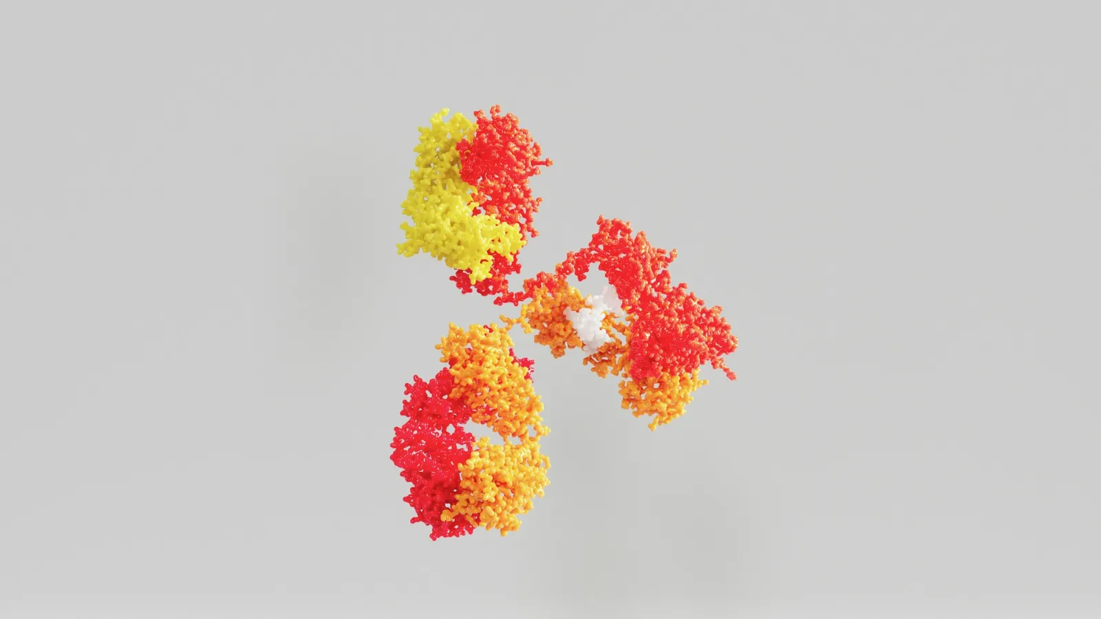
Your NCERT biology book draws an antibody as a Y and writes it as H2L2: two heavy chains and two light chains. That simple picture took more than a decade of patient chemistry to establish, and it came from two scientists working on opposite sides of the Atlantic who attacked the same molecule in different ways. This post tells how Rodney Porter and Gerald Edelman did it, using Nobel Prize records and Porter's Nobel lecture, and checks their numbers with a short worked example.
Who were Porter and Edelman?
Rodney Robert Porter was born on 8 October 1917 in Newton-le-Willows, England, and died on 6 September 1985, according to his Nobel facts page. His Nobel biography says he took a biochemistry degree at Liverpool in 1939, served in the army from 1940 to 1946, including in North Africa and Italy, and then earned a PhD at Cambridge in 1948 under Fred Sanger. He worked at the National Institute for Medical Research from 1949 and became a professor at Oxford in 1967.
Gerald Maurice Edelman was born on 1 July 1929 in New York and died on 17 May 2014, as his Nobel facts page records. His Nobel biography says he trained as a doctor, served as an army physician in Paris, and joined the Rockefeller Institute in New York in 1957, earning his PhD there in 1960.
Why were antibodies so hard to study?
The 1972 Nobel press release says that until 1959 knowledge of what antibodies are was "very vague and incomplete, in spite of a century of research". They are giant molecules, too big for the chemical methods of the time. Both scientists decided to split them into smaller, well-defined pieces.
What did Porter find by cutting with papain?
Porter used papain, a protein-cutting enzyme, on rabbit antibodies. His paper in the Biochemical Journal in 1959 reported the method. In his Nobel lecture he describes what happened. The molecule split into three pieces of similar size, each about one third of the whole. Two were alike and could still bind the antigen. The third could not, and it crystallised easily into diamond-shaped plates.
That last result was so unexpected that Porter's team dismissed the crystals as coming from amino acids rather than protein and threw them away for several months. Only when he showed them to his neighbour, the X-ray crystallographer Olga Kennard, did he learn they were protein crystals. The binding pieces are now called Fab and the crystallising piece Fc.
What did Edelman find by breaking chains apart?
Edelman reasoned that an antibody, like many proteins, might be several chains held together by sulfur bridges. The press release explains that he broke those cross-links and separated the molecule into chains, which on their own did not bind antigen. He reported the first results in 1959 in a short paper titled Dissociation of γ-globulin.
Later, both groups showed the molecule has two light chains and two heavy chains about twice as long. Porter built the model: a Y whose two arms are each one light chain plus the front part of a heavy chain, with the stem made of the rest of the two heavy chains, all held by sulfur links. The binding sites are at the tips of the arms. In 1969 Edelman's group reported the complete amino acid sequence of an entire human antibody molecule in PNAS.
The 1972 Nobel Prize in Physiology or Medicine went to both men "for their discoveries concerning the chemical structure of antibodies".
Worked example: do the masses add up?
Porter's lecture gives rounded molecular weights: about 150,000 for the whole rabbit antibody, about 50,000 for a heavy chain and about 20,000 for a light chain. Let us check whether the pieces fit.
Three papain fragments, each about one third: 150,000 ÷ 3 = 50,000 each.
Four chains: 2 × 50,000 + 2 × 20,000 = 100,000 + 40,000 = 140,000.
Difference from the whole: 150,000 − 140,000 = 10,000, which is 10,000 ÷ 150,000 ≈ 0.067, or about 7%.
One Fab arm is a light chain plus about half a heavy chain: 20,000 + 25,000 = 45,000, close to the 50,000 estimated for each fragment.
The numbers agree to within the rough precision of the 1950s and 1960s measurements. That is how you test a model: if the parts had added to 90,000 or 250,000, the Y would be wrong.
What mistakes do students make with antibody structure?
Reading H2L2 as four identical chains. Two are heavy and two are light, and each pair is identical.
Putting the binding site on the stem. Antigen binds at the tips of the arms, where light and heavy chains meet.
Confusing structure with diversity. How the body makes so many different tips is a separate story, told in our post on Susumu Tonegawa and antibody diversity.
The NCERT Class 12 chapter Human Health and Disease gives the H2L2 picture you need for exams. If protein chains and disulfide bonds feel abstract, the Fermi Concept Library covers every concept in physics, chemistry, biology, mathematics and computing, so you can go back to protein structure before returning to antibodies.
What can a student take from how they worked?
Attack a hard problem from two sides. An enzyme cut and a chemical cut gave complementary clues.
Check that the parts add up. A model is only convincing when the pieces account for the whole.
Do not discard what surprises you. The crystals that looked like a mistake were the key result.
Next step: draw the Y-shaped antibody from memory, label heavy chains, light chains, Fab, Fc and the antigen-binding sites, then check it against the NCERT figure.