

On this page
For centuries, doctors observed patients suffering from "dropsy," a condition marked by severe, generalized swelling. It was often thought to be a primary disease or a sign of heart or liver failure. It was not until 1827 that the English physician Richard Bright connected this swelling to its true origin: the kidneys.
Bright's work laid the foundation for nephrology. Today, the mechanics of how healthy kidneys filter blood—and why protein in the urine is a warning sign—remains a core topic in the Class 11 biology syllabus under "Excretory Products and their Elimination."
How did Richard Bright discover kidney disease?
In his seminal 1827 publication, Reports of Medical Cases, Bright detailed his study of patients with dropsy. He performed post-mortem examinations and consistently found that these patients had damaged, granulated kidneys. To confirm his clinical suspicions while patients were still alive, Bright employed a simple chemical test.
He took a sample of the patient's urine in a spoon and heated it over a candle flame. If the urine turned cloudy and coagulated, it indicated the presence of albumin, a key blood protein. This albuminuria, Bright realised, was a hallmark of kidney disease. His collaboration with the chemist John Bostock confirmed that as albumin was lost in the urine, it became depleted in the blood. For over a century, the triad of dropsy, albuminuria, and kidney damage was known simply as "Bright’s Disease". By linking a physical observation at the bedside with post-mortem anatomical findings, Bright revolutionised diagnostic medicine and established that the kidneys were responsible for filtering blood.
Why do healthy kidneys hold back protein?
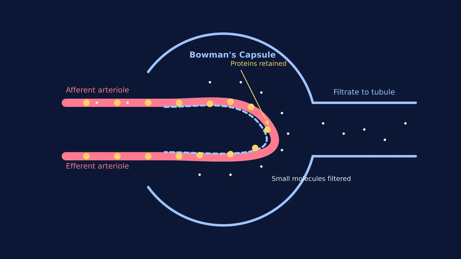
Bright’s discovery makes immediate sense when you look at the structure of the kidney as taught in Class 11 biology. The first step of urine formation is glomerular filtration, which takes place in the Malpighian body (the glomerulus enclosed by Bowman's capsule).
Blood enters the glomerulus under pressure from the afferent arteriole. The filtration membrane has three layers: the endothelium of the blood vessels, the basement membrane, and the epithelium of Bowman's capsule. The epithelial cells, known as podocytes, are arranged to leave tiny spaces called filtration slits.
This process is called ultrafiltration because it is extremely fine. Almost all constituents of blood plasma pass into the capsule to form the filtrate, except for large molecules like plasma proteins. Because proteins are too large to pass through the healthy filtration slits, the resulting filtrate is essentially protein-free plasma. After this initial filtration, the fluid travels through the rest of the nephron, including the loop that concentrates urine, before its final concentration is adjusted by hormones regulating kidney function.
When the filtration membrane is damaged—as Bright observed in his patients—proteins leak through, leading to proteinuria. The resulting loss of albumin lowers the osmotic pressure of the blood, which causes fluid to leak into tissues and creates the severe swelling, or dropsy, that Bright's patients suffered from.
How to prepare for biology exams on excretion?
When studying the excretory system for board exams or NEET, understanding why certain molecules pass into the filtrate while others do not is crucial. If you have gaps in your understanding of human physiology, the Fermi Concept Library maps out every concept in biology, physics and chemistry, each with its own lesson.
Once you understand the theory of ultrafiltration, you have to apply it to questions about the glomerular filtration rate (GFR) and the composition of the filtrate. You must be able to trace the path of molecules as they move from the blood into Bowman's capsule and follow the filtrate as it travels through the proximal and distal tubules.
If you are stuck on a question, the Fermi Problem Library offers worked problems with a step-by-step solver that explains the reasoning rather than just printing an answer.
Richard Bright's candle-flame test was a leap forward in medical diagnostics. By linking a visible symptom like swelling and a chemical sign like protein in urine to a specific organ, he showed that disease could be understood through careful observation of the body's filters. His work transformed the kidney from a mysterious organ into one that could be clinically monitored, paving the way for modern medical science.


