

On this page
In 1981, a team at the University of Cambridge published a paper in *Nature* that gave scientists a new way to study genetics and human disease. Sir Martin Evans and Matthew Kaufman had taken early mouse embryos and isolated cells from them that could grow indefinitely in a petri dish. Crucially, these cells were pluripotent: they had the potential to turn into any type of tissue in the adult body.
Until then, maintaining cells in this flexible, unspecialised state in a laboratory had been nearly impossible. By finding the right conditions to keep them dividing without forcing them to specialise, Evans made genetic engineering possible in mammals. In 2007, he shared the Nobel Prize in Physiology or Medicine for this work.
What is an embryonic stem cell?
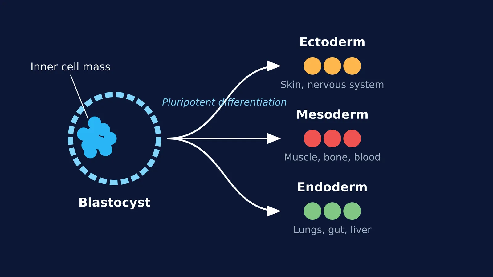
A few days after fertilisation, a mammalian embryo forms a hollow ball of cells called a blastocyst. The blastocyst has two main parts: an outer layer that will become the placenta, and a cluster of cells inside called the inner cell mass.
The cells of the inner cell mass are embryonic stem (ES) cells. They are described as pluripotent because they can differentiate into all three primary germ layers—the ectoderm, mesoderm, and endoderm—which go on to form every organ and tissue in the body. If you are studying embryonic development for NEET, understanding the sequence from zygote to morula and then to blastocyst is a core requirement.
If you find it difficult to picture how these cell layers fold and migrate during early development, the Fermi Concept Library contains lessons that break down embryogenesis step by step, allowing you to check your understanding as you go.
How did Evans culture them in 1981?
The challenge before 1981 was that when scientists tried to grow cells from early embryos, the cells would rapidly differentiate into specific tissue types or simply stop dividing.
Evans and Kaufman worked with the 129 strain of mice. They took blastocysts, extracted the inner cell mass, and placed the cells in culture dishes. By carefully controlling the environment, they established stable cell lines that continued to divide while retaining their pluripotency. The cells could be frozen, thawed, and multiplied, providing a constant supply of unspecialised cells for research.
Why make knockout mice?
The value of embryonic stem cells became clear when Evans realised they could be used as a delivery vehicle for genetic changes.
Scientists could take an ES cell in a dish and intentionally modify its DNA—for example, by "knocking out" or disabling a specific gene. They could then inject this modified ES cell into a normal mouse blastocyst and implant it into a surrogate mother. The resulting mouse, called a chimera, would have some tissues made from the modified cells. By breeding these mice, scientists could produce offspring in which every cell lacked the target gene.
This technique led to the creation of knockout mice. By observing what goes wrong when a specific gene is missing, researchers have been able to model human conditions such as cystic fibrosis, cardiovascular disease, and cancer, testing new treatments safely before they reach human trials.
If you are working through genetics questions and struggling to predict the outcomes of genetic crosses or mutations, the Fermi Problem Library has a step-by-step solver that explains the reasoning behind heredity problems rather than just printing an answer.
How can you trace cell lineages in your studies?
When learning about stem cells and human reproduction, students often mix up which germ layer produces which organ. Use this simple mapping strategy:
Ectoderm (outside): Think of things that interact with the outside world. It forms the skin (epidermis) and the entire nervous system (brain, spinal cord, nerves).
Mesoderm (middle): Think of the body's structural and transport systems. It forms muscles, bones, the heart, blood vessels, and kidneys.
Endoderm (inside): Think of the internal linings. It forms the lining of the digestive tract, the lungs, and associated organs like the liver and pancreas.
By linking Evans's 1981 discovery of pluripotent cells to the germ layers they eventually form, you can see how a single cluster of cells in a dish holds the blueprint for an entire organism. Read more about the early stages of human development in our guide to fertilisation to implantation, or explore another milestone in biotechnology: recombinant insulin production.


