

On this page
In 1906, the beating of the heart was known to be an electrical process, but the exact starting point of the signal remained a mystery. Earlier that year, the Japanese pathologist Sunao Tawara and the atrioventricular node of the heart had mapped the AV node and the bundle of His, which carried the impulse to the ventricles. But what gave the AV node its signal?
The answer came from a makeshift laboratory at a farmhouse in Kent, England. Sir Arthur Keith, a Scottish anatomist, and Martin Flack, a medical student, decided to search for the origin of the heartbeat.
Who discovered the SA node pacemaker?
The discovery belonged to Arthur Keith and Martin Flack. Keith was an established anatomist, while Flack was a medical student assisting him (Wikipedia).
During the summer of 1906, they systematically examined the hearts of various mammals. While Keith was out on a bicycle ride, Flack examined microscopic sections of a mole's heart. He noticed a distinct, specialised cluster of muscle fibres that looked different from the surrounding tissue.
When Keith returned, they confirmed the finding across other vertebrate hearts, including dogs and humans. They originally named this structure the "sino-auricular node" and published their findings in the Journal of Anatomy and Physiology in April 1907 (Keith & Flack, 1907).
Where is the sinoatrial node located?
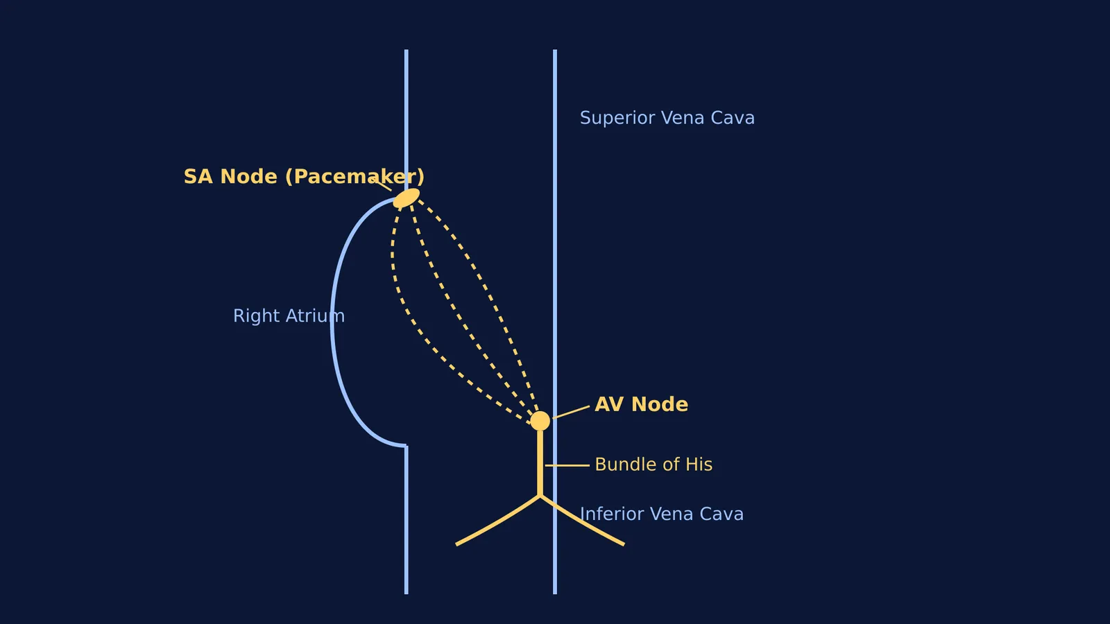
Keith and Flack found the SA node exactly where the superior vena cava—the large vein carrying deoxygenated blood from the upper body—enters the right atrium of the heart.
They noted that this small knot of cells was richly supplied with nerve fibres. Because of its unique structure and strategic location, they correctly concluded that it was the dominant rhythm-maker, acting as the heart's natural pacemaker.
If you need to visualise these structures for your biology studies, the Fermi Concept Library holds every concept in physics, chemistry, biology, mathematics and computing, each with its own lesson and its place in the map.
How did they find it in a mole's heart?
The choice of animals was crucial to their discovery. Keith and Flack used comparative anatomy to look for structures that remained consistent across different species. The mole's heart provided exceptionally clear tissue sections, making the distinct, delicate network of muscle fibres of the SA node stand out under the microscope.
They observed that this specialised node of tissue was actually a remnant of the sinus venosus, a chamber found in the primitive hearts of fish and amphibians. In mammals, this chamber has been absorbed into the wall of the right atrium during embryonic development. By tracing this evolutionary history, they pinpointed where the heartbeat begins.
Their work proved that the SA node is an evolutionary feature conserved across vertebrates, ensuring that the heart contracts in a coordinated, top-down sequence. Decades later, understanding this electrical pathway would make breakthroughs like Christiaan Barnard and the first human heart transplant possible, as surgeons had to know exactly how to restart a transplanted heart.
How does the heart's electrical pathway work?
Understanding the SA node is easier when you see it as the first step in a cascade. Here is the sequence of a normal heartbeat:
SA Node (Sinoatrial Node): Fires the initial electrical impulse, causing the left and right atria to contract.
AV Node (Atrioventricular Node): Catches the signal and delays it slightly, allowing the ventricles to fill with blood.
Bundle of His: Carries the impulse down the septum between the ventricles.
Purkinje Fibres: Spread the signal through the ventricular walls, forcing them to pump blood out of the heart.
If you are stuck on understanding how these impulses coordinate, Fermi's AI tutor can work out what you already know, build a plan for the session, and teach through it by drawing the figure and checking understanding as it goes.


