
On this page
Most viruses are far too small to see with the microscope in your school lab. Before modern genetic tests, the way to know a virus was there was often to see it with an electron microscope, and few people were better at that than June Almeida. Her pictures helped identify new viruses, including some of the first human coronaviruses. This post follows her BMJ obituary and her own papers, works through the physics of why electrons can show what light cannot, and ends with what students can learn from her.
Who was June Almeida?
Her obituary in the BMJ records that she was born on 5 October 1930 and grew up in a tenement flat in Glasgow. She did well at school but left at 16, in 1947, without the money to go to university. She became a laboratory technician in histopathology at Glasgow Royal Infirmary, then worked at St Bartholomew's Hospital in London.
After she married and moved to Canada, a job came up for an electron microscopy technician at the Ontario Cancer Institute in Toronto. With no formal qualifications, she co-authored many papers on virus structure. In 1964 Professor A. P. Waterson persuaded her to join him in London, and her publications later earned her a DSc, a higher doctorate.
What did she find in 1967?
David Tyrrell, director of the Common Cold Research Unit, had grown cold viruses that would not grow by normal methods, using organ cultures: small pieces of living airway tissue kept alive in the lab. In 1965 he and M. L. Bynoe reported a new type of common-cold virus grown this way. The next question was what such viruses looked like.
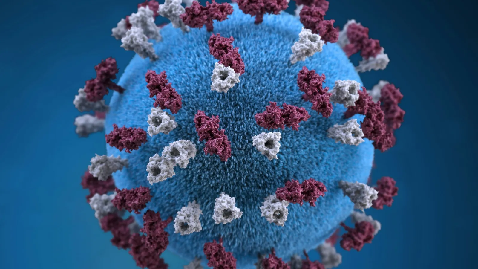
Almeida and Tyrrell's 1967 paper in the Journal of General Virology described "a simple method" for examining organ cultures by electron microscopy for virus particles. With it they found three viruses that had not been described before. Two had particles resembling those of infectious bronchitis, a virus of chickens, and the third resembled the parainfluenza viruses.
In 1968 a short note in Nature, titled Coronaviruses, grouped viruses of this kind under that new name. The BMJ obituary notes that the more recently recognised SARS virus belongs to the same group.
Why can electrons show what light cannot? A worked example
The detail any microscope can show is limited by the wavelength of what it uses. The NCERT Class 12 physics chapter on ray optics gives visible light a wavelength of about 400 nm to 750 nm. The NCERT chapter Dual Nature of Radiation and Matter gives the de Broglie relation for a moving particle: λ = h/p.
Find the wavelength of an electron accelerated from rest through 100 V.
Kinetic energy: K = eV = (1.6 × 10⁻¹⁹ C)(100 V) = 1.6 × 10⁻¹⁷ J.
Momentum: p = √(2mK) = √(2 × 9.11 × 10⁻³¹ kg × 1.6 × 10⁻¹⁷ J) = √(2.92 × 10⁻⁴⁷) ≈ 5.40 × 10⁻²⁴ kg m/s.
Wavelength: λ = h/p = (6.63 × 10⁻³⁴ J s) ÷ (5.40 × 10⁻²⁴ kg m/s) ≈ 1.23 × 10⁻¹⁰ m = 0.123 nm.
Compare with violet light: 400 nm ÷ 0.123 nm ≈ 3,300.
So even a 100 V electron has a wavelength about 3,300 times shorter than violet light. Electron microscopes use much higher voltages, which make the wavelength shorter still. In practice their detail is limited by the magnetic lenses rather than by the wavelength.
Common mistakes:
Using V in place of eV. K = eV in joules. Multiply by the electron's charge.
Forgetting the square root. p = √(2mK), not 2mK.
Mixing nm and m. 1 nm = 10⁻⁹ m, so 1.23 × 10⁻¹⁰ m is 0.123 nm.
Our post on de Broglie wavelength problems has more practice, and the story of the instrument itself is in our post on Ernst Ruska.
What else did her methods reveal?
The BMJ obituary describes her technique of immune electron microscopy. She mixed a virus sample with antibodies, which made the virus particles clump together so they were easier to find and identify. With it she was the first to see rubella virus, which had been known to harm babies in pregnancy for more than 25 years.
She also showed that the hepatitis B virus has two distinct parts, one on the surface and one inside. Antibodies to the surface part protect against infection, and that surface part is what current hepatitis B vaccines stimulate. Our post on Baruch Blumberg and hepatitis B tells the rest of that story.
She taught the method to Albert Kapikian of the US National Institutes of Health, who used it to identify the "Norwalk agent", now called norovirus. Her negative staining methods let virologists identify many infections within minutes of collecting a sample.
She later worked at the Wellcome Research Laboratories on diagnostic tests and vaccines, retired in 1985, and in the late 1980s returned to St Thomas' Hospital as an adviser, helping publish some of the first high-quality electron micrographs of HIV. She died on 1 December 2007.
What can a student take from Almeida's way of working?
A late or unusual start is not the end. She left school at 16 and still earned a doctorate through published work.
Master one tool deeply. Her skill with the electron microscope made her the person others came to.
Prefer simple methods. The obituary credits her success to finding simple explanations and using few reagents.
Teach what you know. Kapikian's norovirus work came from a method she taught him.
If wave-particle duality still feels strange, Fermi's Simulation Lab has interactive simulations for ideas that are easier to see than read.
Next step: repeat the worked example for an electron accelerated through 400 V and check that the wavelength halves.