

On this page
In the early 1950s, doctors treating patients with mitral valve disease faced a serious problem. A narrowed valve required surgery, but a leaking valve did not. Yet surgeons had no safe, non-invasive way to tell the difference before they opened the chest. Swedish cardiologist Inge Edler at Lund University wanted a way to "see" the heart's movement from the outside.
He asked physicist Hellmuth Hertz—the son of Nobel laureate Gustav Hertz and grandnephew of Heinrich Hertz—if radar might work. Hertz knew radar waves would not pass through the body well, but he suggested high-frequency sound instead. Industry was already using ultrasound to find cracks in metal.
How did a shipyard flaw detector help invent echocardiography?
In May 1953, Hertz visited Tekniska Rontgencentralen AB at the Kockum's shipbuilding yard in Malmö, Sweden. They used a Siemens ultrasonic reflectoscope to test steel hulls for microscopic flaws. Hertz pressed the industrial transducer to his own chest and watched the oscilloscope screen. He saw a signal pulsating in time with his heartbeat, proving that ultrasound could reflect off the interfaces between blood and heart tissue. He borrowed the heavy machine for the weekend to test at the cardiac laboratory at Lund University.
Seeing a physical principle in action changes how you understand it. If you are studying how waves reflect at boundaries, the Fermi Simulation Lab has interactive simulations for the ideas that are easier to see than to read.
What was the first echocardiogram in 1953?
To continue their work, Hertz secured a year-long loan of a medical ultrasound reflectoscope (the "Ultraschall Impulsgerät") from Siemens. On October 29, 1953, they placed the ultrasonic probe over a human heart and recorded the first moving pictures.
They observed an echo moving back and forth along the X-axis of the oscilloscope screen at a depth of 8 to 9 cm from the chest wall. The sound waves were bouncing off the structures inside the heart, and the time it took for the echo to return revealed their exact depth. This inaugurated the field of ultrasound cardiography.
How do you calculate tissue depth from an ultrasound echo?
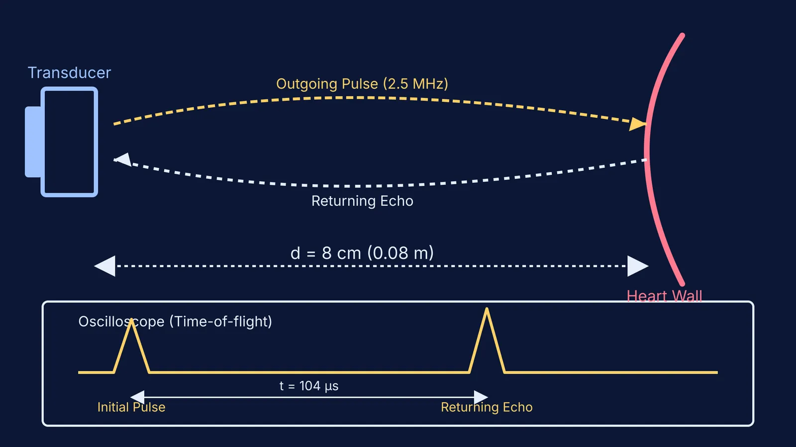
The reflectoscope measured the time (t) it took for the sound pulse to travel to the heart wall and back. Since the standard assumed speed of sound in human soft tissue (v) is roughly 1540 m/s, the depth (d) of the tissue is found using this simple framework:
d = (v × t) / 2
For an echo returning from a depth of 8 cm (0.08 m), the round-trip time is:
t = (2 × 0.08) / 1540 = 0.000104 seconds, or 104 microseconds.
This time-of-flight principle is still how modern ultrasound machines map the body. Complex medical machines are built on foundational physics. When you need to master those underlying rules, Fermi's AI tutor can work out what you already know and build a plan for the session, so you sit down to a clear next step.
Why is Inge Edler called the Father of Echocardiography?
Edler developed the M-mode (time-motion) technique to capture these fleeting echoes. By pulling photographic film past the oscilloscope screen at a constant speed, he turned the back-and-forth blip into a continuous wavy line. This allowed doctors to measure the exact thickness and movement of the mitral valve without making a single incision.
For his pioneering work, Edler is recognised as the "Father of Echocardiography," and he shared the 1977 Lasker-DeBakey Clinical Medical Research Award with Hertz. Today, the technique they proved with a shipyard tester is one of the most vital diagnostic tools in cardiology.


