On this page
Cellular respiration is the process by which living things extract energy from food, a mechanism that relies on a chain of electron carriers. The discovery of these carriers, known as cytochromes, was made in 1925 by David Keilin at the Molteno Institute in Cambridge.
Keilin’s work not only solved the mystery of how cells use oxygen but also validated a dismissed discovery from 40 years earlier. His careful observation using spectroscopy bridged the gap between cellular structure and chemical function, forming the basis of modern bioenergetics.
Who discovered cytochromes?
The story of cytochromes begins in 1884 with Charles Alexander MacMunn, an Irish physician who observed light-absorption bands in animal tissues source. MacMunn named these pigments "myohaematin" and "histohaematin" and proposed that they acted as respiratory catalysts.
MacMunn was a practitioner working in relative isolation, operating an unpaid laboratory from his home. In contrast, Felix Hoppe-Seyler was the leading physiological chemist in Europe, known for having named hemoglobin and discovered its oxygen-binding properties. When MacMunn published his findings, Hoppe-Seyler dismissed them. He argued that the bands were merely decomposition products of hemoglobin caused by the extraction process source. The scientific community sided with the established authority, and unable to defend his work against such a prominent figure, MacMunn abandoned the research. His discovery lay forgotten for decades.
It was David Keilin who vindicated MacMunn. In his 1925 paper, "On cytochrome, a respiratory pigment, common to animals, yeast, and higher plants," Keilin demonstrated that these pigments were present even in organisms without hemoglobin, such as yeast and plants source. He renamed the pigments "cytochromes" (meaning "cell pigments") and proved they were a functional part of the living cell, not a byproduct of decay.
How did David Keilin observe cytochromes?
Keilin made his discovery while studying the life cycle of the horse bot-fly (*Gasterophilus intestinalis*) source. To examine the tissues, he used a Zeiss microspectroscope ocular—a tool that combined a microscope with a spectroscope.
Traditional spectroscopes required samples to be extracted and purified in solutions, a process that risked altering the biological molecules. By attaching a spectroscope directly to a microscope, Keilin bypassed this limitation. He focused light through the intact wing muscles of the living bot-fly directly into the prism of his instrument, splitting the transmitted light to reveal the exact absorption lines of the molecules as they functioned in their natural state.
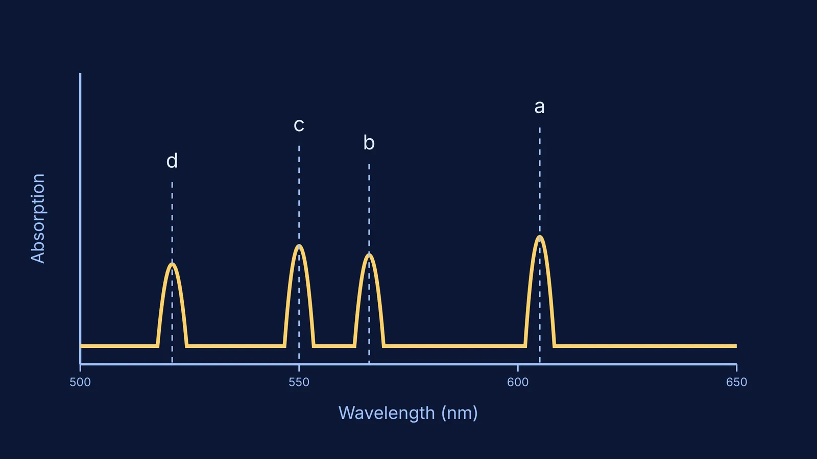
When observing the insect wing muscles, Keilin saw a four-banded absorption spectrum, which he labelled a, b, c, and d source.
Keilin noticed that the spectrum changed based on the cell's activity. When the tissue was oxygenated, the absorption bands vanished. When the oxygen was consumed or a reducing agent was added, the four bands reappeared. This reversible change proved that the cytochromes were actively accepting and passing on electrons, functioning as an intermediary respiratory chain that transfers electrons to oxygen.
Ideas like the electron transport chain can be hard to picture from text alone. In the Fermi Simulation Lab, you can run interactive simulations to see how electrons flow through cytochromes and power the cell. Once you understand the mechanism, you can see how it ties into larger processes, like respiration as an amphibolic pathway where fats and proteins enter the cycle.
How can you analyse historical experiments?
When studying landmark discoveries for competitive exams, you need to understand how the scientist reached their conclusion. Biological systems are complex, and researchers must carefully isolate variables to prove a functional relationship. Use this three-step framework when reviewing historical experiments:
The Observation: What exactly did the scientist see or measure? For Keilin, it was not just the presence of a pigment, but the appearance and disappearance of four specific absorption bands in living tissue.
The Variable: What condition was changed to affect the observation? Keilin introduced or removed oxygen from the environment of the cell. He also introduced chemical reducing agents to artificially replicate the oxygen-deprived state.
The Deduction: How did the observation link to a biological function? The reversible nature of the bands proved the pigment was repeatedly being oxidised and reduced. This meant it was not a static structural component, but an active participant that transferred electrons.
If you find yourself memorising facts without grasping the underlying logic, Fermi's AI tutor can work out what you already know and build a plan for the session, guiding you through the reasoning step by step. This analytical approach will help you tackle complex physiology and biochemistry questions, whether you are studying human respiration or plant mechanisms like photorespiration and RuBisCO.