On this page
In 1759 a young doctor named Caspar Friedrich Wolff argued that embryos do not start as tiny, finished copies of adults. Their organs build up gradually from simpler tissue, and chick embryos became his best evidence. That idea, epigenesis, is one root of the germ layers in your biology textbook.
Who was Caspar Friedrich Wolff?
Wolff was born in Berlin on 18 January 1734 and died in St Petersburg on 22 February 1794, according to the Embryo Project Encyclopedia at Arizona State University. He earned his MD at the University of Halle in 1759 with a dissertation, Theoria Generationis, that described development in both plants and animals as a process involving layers. His name survives in anatomy: the embryonic kidneys he described are called Wolffian bodies, and their ducts are Wolffian ducts.
What did scientists believe before Wolff?
The leading view was preformation, the idea that an organism develops by the unfolding of a form that is somehow present from the outset (Embryo Project: Wolff). The Italian microscopist Marcello Malpighi had looked at chicken eggs early in incubation and reported well-formed organs and structures, and preformationists leaned on his descriptions (Embryo Project: Ovism). Malpighi's sharp eyes are also the subject of our piece on Marcello Malpighi and the capillaries Harvey could not see.
Albrecht von Haller, a strong proponent of preformation, also studied chick embryos under the microscope. He argued that the chick already existed inside the egg and that fertilisation simply set its tiny, transparent heart beating (Embryo Project: Albrecht von Haller). His key evidence was the membrane continuity proof: the yolk seemed to be attached to the embryo's small intestine, so, he reasoned, embryo and yolk must have been made together.
What did Wolff see in chick embryos?
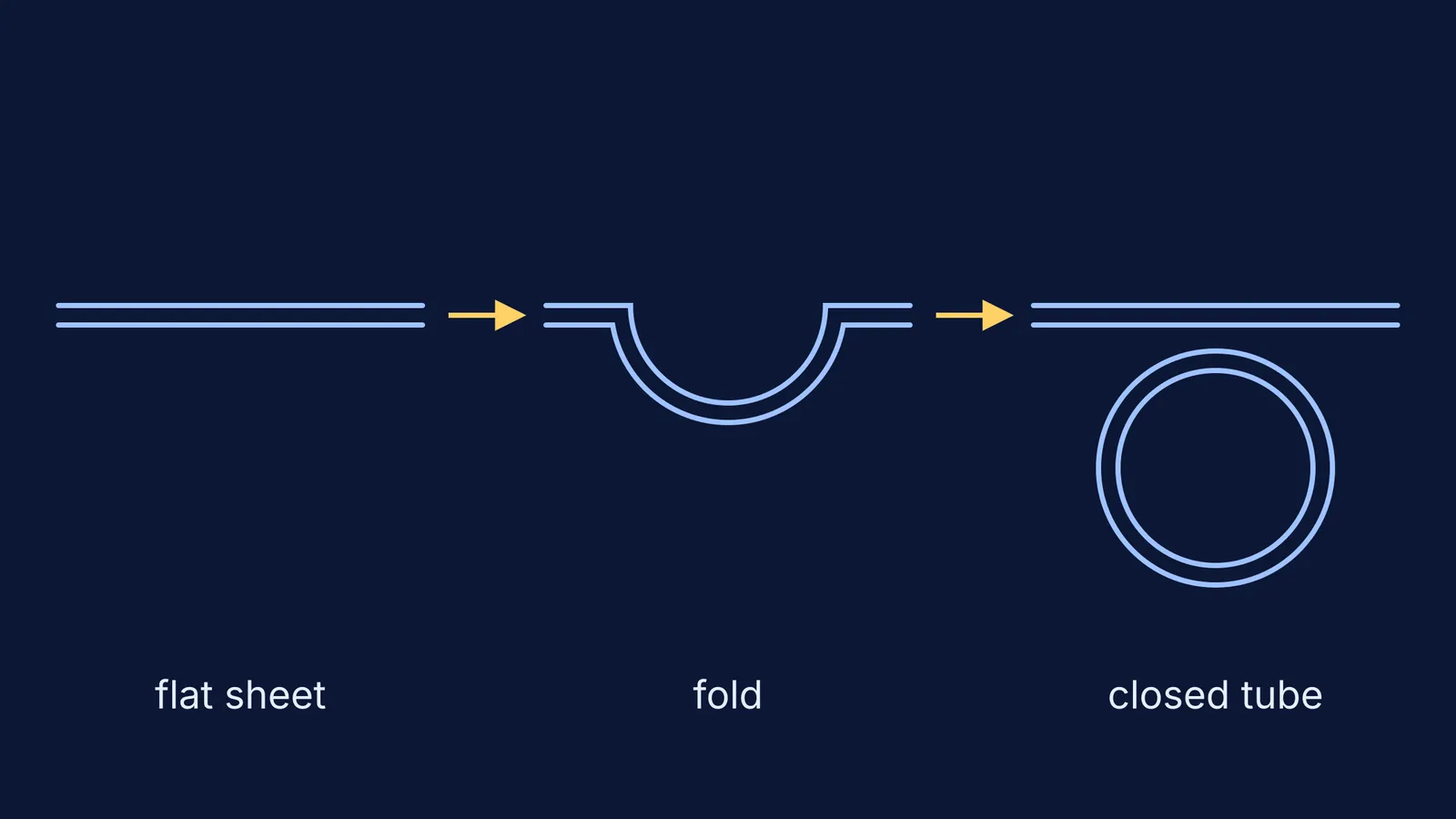
Wolff saw parts that were not there at first and appeared step by step. The clearest case was the gut. In De Formatione Intestinorum, published in 1768, he showed that the chick intestine forms by the folding of tissue that detaches from the embryo's lower (ventral) surface, and that the folds eventually close into a tube (Embryo Project: Needham's A History of Embryology). He argued that this proved the intestine was not preformed and that organs appear gradually.
That also undid Haller's best argument. The Embryo Project notes that Wolff showed the continuity did not exist as Haller had described it, using more skilled microscopy than Haller, who had poor eyesight (Embryo Project: Albrecht von Haller). The publisher of a modern French edition calls the study the first great text of modern embryology, because it describes the embryo's in-between forms (Brepols edition).
Why did it take so long for epigenesis to win?
Haller was far better known than Wolff, and his influence did much to keep preformation alive through the late 1700s (Embryo Project: Needham's A History of Embryology). Wolff's work paved the way for later embryologists, including Karl Ernst von Baer (Embryo Project: Wolff), and historians of embryology see his folding sheets as an early form of germ-layer theory (UNSW Embryology: Caspar Wolff).
You use that idea in Class 11. The NCERT Biology chapter on the Animal Kingdom defines triploblastic animals as those whose developing embryo has three germinal layers: ectoderm, mesoderm and endoderm (NCERT Class 11 Biology, Chapter 4). Much later, the question moved from what forms to what tells tissue what to become; that story is in Hans Spemann, Hilde Mangold and the embryo organizer.
How can you study embryology the way Wolff did?
Wolff's real tool was a question: what does each idea predict, and what do I actually see, stage by stage? Here is the gut worked through.
Write both predictions. Preformation: a tiny gut is there from the start and only grows. Epigenesis: no gut at first, then a flat sheet, a groove, a tube.
List the stages you would check: flat sheet, fold, closed tube.
Ask which observation one idea cannot explain. A stage with a fold but no tube makes no sense if a finished gut was there all along.
Draw the three stages from memory, then write one verb under each arrow, such as fold or close.
The same routine works for any development topic, from germ layers to fertilisation and implantation in the first week of pregnancy. Before you call a diagram learnt, check:
What exists at the first stage?
What is new at the next stage, and where did it come from?
Which verb describes the change: fold, split, fuse or grow?
If a term such as mesoderm keeps slipping, the Fermi Concept Library gives every concept in biology its own lesson and its place in the map. If you are not sure where to begin with a whole chapter, Fermi's AI tutor can work out what you already know and build a plan for the session.
Wolff's lesson still holds: look again, stage by stage.