On this page
In 1980, solving the atomic structure of the ribosome was widely considered an impossible problem by the scientific establishment. The ribosome is the cell's essential protein factory, responsible for reading genetic instructions encoded in mRNA and assembling amino acids into the proteins that keep every living organism alive. But to truly understand how this microscopic machine works, scientists needed to map the exact position of hundreds of thousands of atoms using a technique known as X-ray crystallography.
To do this, researchers first had to grow a highly ordered crystal made entirely of pure ribosomes. Because ribosomes are massive, flexible, and chemically unstable, the scientific consensus held that they could never form the rigid, perfectly aligned lattice that a crystal requires. Ada Yonath chose to ignore this consensus, embarking on a remarkable research journey that would take over two decades and fundamentally transform structural biology.
Why is the ribosome structure hard to map?
X-ray crystallography works by firing a focused beam of X-rays at a crystal. The atoms in the crystal scatter the X-rays, producing a complex diffraction pattern that computers can use to calculate the precise three-dimensional structure of the molecule. The better and more ordered the crystal, the sharper and more accurate the resulting picture.
Yonath worked for over two decades to grow high-quality crystals of ribosomes. The vast majority of these early experiments failed. When she finally obtained usable crystals, they proved to be incredibly fragile. The intense X-ray beam required for the imaging process systematically destroyed them, melting the crystal before a complete diffraction pattern could be recorded. She urgently needed ribosomes that were tough enough to survive the rigorous imaging process without falling apart.
How did Ada Yonath use extremophiles and cryocrystallography?
Drawing inspiration from an article about polar bears that pack their ribosomes tightly during winter hibernation, Yonath began to look for bacteria that naturally thrive in harsh environments. She reasoned that organisms surviving under extreme stress would have evolved sturdier cellular machinery. She extracted ribosomes from hardy bacteria living in extreme environments like the Dead Sea and thermal springs. Because these extremophile bacteria are adapted to function in severe conditions, their ribosomes are naturally robust and far more stable during the crystallization process.
To overcome the persistent problem of radiation damage from the X-ray beam, Yonath developed a technique called cryocrystallography. By flash-freezing the delicate ribosome crystals in liquid nitrogen to −185°C, she discovered that they could withstand the X-ray bombardment long enough to yield clear data. This crucial breakthrough is now a standard tool used in structural biology laboratories worldwide.
In 2000, after twenty years of persistent effort, her team published the first complete atomic maps of both the small and large ribosomal subunits. Her unyielding dedication to mapping the ribosome earned her the 2009 Nobel Prize in Chemistry.
If you have gaps in your understanding of transcription and translation, the Fermi Concept Library breaks down the entire process step by step. Because molecular machinery is incredibly hard to picture from text alone, the Fermi Simulation Lab offers interactive simulations for ideas that are easier to see than to read, such as how a ribosome chemically reads RNA.
What does the ribosome structure reveal about antibiotics?
Many of the most critical antibiotics we rely on today, such as erythromycin and tetracycline, cure bacterial infections by specifically shutting down bacterial ribosomes while leaving our own human ribosomes completely unaffected. Before Yonath's crystallographic work, scientists knew that these life-saving drugs worked, but they did not know exactly *how* they managed to halt the protein assembly line.
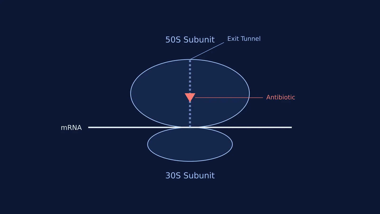
Her detailed maps revealed the precise atomic pockets where over two dozen antibiotics selectively bind to the bacterial ribosome. For example, some drugs act by plugging the polypeptide exit tunnel like a cork, physically blocking the new protein from leaving the ribosome. This intricate structural blueprint now allows modern chemists to intelligently design entirely new drugs to combat the growing global crisis of antibiotic resistance.
You can read more about how cells protect their genetic instructions in our article on Aziz Sancar and how cells repair damaged DNA, or trace the fascinating history of protein mapping back to Margaret Dayhoff and the first protein sequence database.
What can we learn from her approach to an impossible problem?
Yonath's journey from being dismissed as an unrealistic dreamer by her peers to eventually winning a Nobel Prize offers a powerful masterclass in long-term problem solving:
Find a robust model: When a standard experimental system breaks down under stress, look to nature for alternatives. Yonath elegantly solved the stability problem by studying extremophiles.
Build the missing tools: If the existing methods consistently destroy your sample, you have to invent entirely new ones. Cryocrystallography was a necessary and highly inventive detour required to reach the final goal.
Expect a long feedback loop: Success is rarely immediate in fundamental research. Yonath worked for over two decades before obtaining her definitive high-quality crystal structures.
Ignore the consensus: A problem broadly labelled as impossible by the prevailing field is often just a problem waiting for a fresh, unconventional approach.