

On this page
The wall of the human uterus is a highly dynamic and resilient structure. It must be strong enough to protect a developing fetus, elastic enough to expand dramatically during gestation, and active enough to shed its inner lining during each menstrual cycle. To accomplish these vastly different biological tasks, the uterine wall relies on three specialised tissue layers: the outer perimetrium, the middle myometrium and the inner endometrium. Understanding how these layers behave during menstruation, pregnancy and childbirth is fundamental to human reproductive biology.
What are the layers of the uterus?
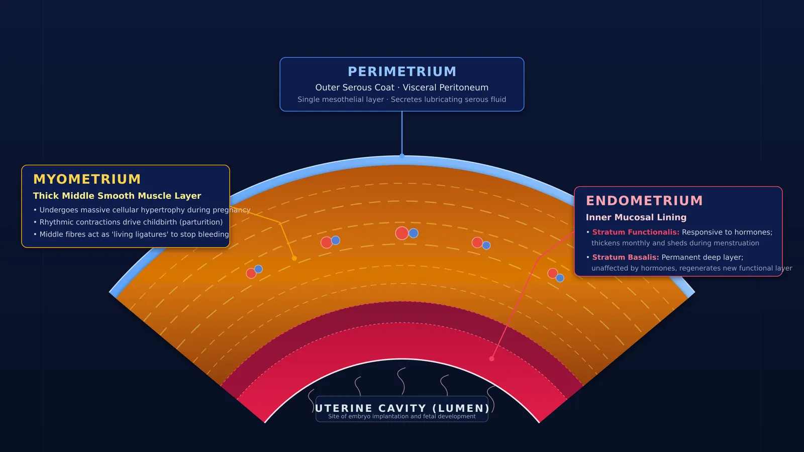
If you examine a cross-section of the uterine wall under a microscope, you will find three distinct layers working in harmony, each with a specialised biological function and tissue composition:
Perimetrium: The thin, outermost serous layer that protects the uterus from mechanical friction against neighbouring pelvic organs.
Myometrium: The thick middle layer composed of smooth muscle, responsible for structural stretching during pregnancy and forceful contractions during childbirth.
Endometrium: The inner mucosal lining that thickens each month to receive a fertilised egg and sheds during menstruation if fertilization does not occur.
These layers form the foundation of human reproductive physiology. To see where uterine anatomy fits into human reproduction, each concept has its own lesson and its place in the map within the Fermi Concept Library.
What happens to the endometrium during the menstrual cycle?
The endometrium is the most biologically dynamic layer of the uterine wall. It directly borders the uterine cavity and is divided into two functionally distinct sub-layers: the permanent stratum basalis (basal layer) and the temporary stratum functionalis (functional layer).
During the proliferative phase of the menstrual cycle, rising oestrogen levels produced by ovarian follicles stimulate the stratum functionalis to undergo rapid cellular growth. According to a StatPearls review of menstrual cycle physiology, the endometrial lining typically reaches a thickness of 8 to 12 mm by the end of the proliferative phase. This thick, highly vascular and nutrient-rich lining prepares the uterus to support a fertilised egg through the first week of pregnancy.
If an embryo does not implant, progesterone levels drop sharply. This withdrawal of hormones causes the specialized spiral arteries within the stratum functionalis to constrict, starving the superficial tissue of oxygen and nutrients. The functional layer rapidly breaks down and sloughs off, resulting in menstrual flow. Once bleeding stops, the deeper stratum basalis—which is unaffected by the hormone drop and is never shed—acts as a regenerative base, growing a brand new functional layer for the subsequent cycle.
How does the myometrium adapt during pregnancy and birth?
The myometrium is by far the thickest part of the uterine wall. Because it consists of dense, interwoven bundles of smooth muscle fibres, it is built to withstand and drive extreme mechanical changes.
During pregnancy, the myometrium undergoes massive physical growth to accommodate the developing fetus. A StatPearls overview of pregnancy physiology notes that a non-pregnant uterus weighs approximately 70 grams and has an internal resting volume of just 10 mL. By the end of a full-term pregnancy, the uterus increases in weight to approximately 1,100 grams and its volume capacity expands to 5,000 mL. This dramatic expansion is powered primarily by hypertrophy, a process where existing individual smooth muscle cells increase massively in size rather than multiplying in number.
According to anatomical descriptions of the myometrium, the smooth muscle is organised into three interconnected layers:
Inner layer (sub-endometrial): Composed of longitudinal and circular fibres near the endometrium, this zone assists in subtle wave-like peristaltic contractions that facilitate sperm transport and early embryo orientation.
Middle layer: The thickest layer, consisting of a dense, mesh-like network of crisscrossing and circular fibres intertwined with arcuate and radial blood vessels.
Outer layer: Formed of longitudinal muscle fibres that provide overall tensile strength and merge directly with the supporting pelvic ligaments.
During childbirth, these muscle fibres contract powerfully and rhythmically to expel the baby, a process driven by the hormones behind birth and milk. Immediately after the placenta detaches from the uterine wall, the crisscrossing muscle fibres of the middle layer perform a critical final task: they act as "living ligatures." By clamping down tightly on the severed blood vessels that supplied the placenta, they mechanically shut off the flow of blood, preventing postpartum haemorrhage.
What is the role of the perimetrium?
The perimetrium acts as the outermost protective coat of the uterus. As detailed in the StatPearls review of uterine anatomy, it is a serous membrane formed by a single layer of squamous mesothelial cells resting on a thin bed of loose connective tissue.
Because the perimetrium is continuous with the visceral peritoneum, it extends over the fundus and the body of the uterus before folding onto neighbouring pelvic organs, forming anatomical recesses such as the pouch of Douglas between the uterus and the rectum. The smooth mesothelial surface secretes serous fluid that lubricates the exterior of the organ. This lubrication allows the uterus to shift, expand and contract smoothly within the pelvis without causing painful friction against the bladder and intestines.


