

On this page
How do the placenta and umbilical cord exchange materials without mixing blood?
During human pregnancy, maternal and foetal bloodstreams never merge. If they mixed directly, the high pressure of the maternal arterial system would instantly rupture fragile foetal capillaries. Furthermore, because a foetus expresses paternal antigens, direct contact with maternal lymphocytes would trigger immune rejection.
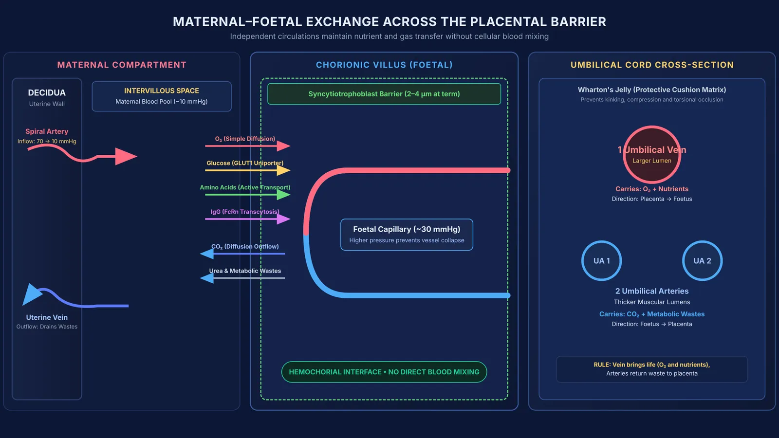
The human body solves this through a hemochorial placenta. As established in Vascular Biology of the Placenta, maternal blood enters the intervillous space from spiral arteries at approximately 10 mmHg, bathing foetal chorionic villi. Inside these villi, foetal capillaries maintain an internal pressure of approximately 30 mmHg, preventing their collapse under surrounding maternal blood. Blastocyst attachment is explored in From fertilisation to implantation.
If you find it challenging to visualize how pressure balances prevent fluid mixing while facilitating transport, the Fermi Concept Library covers every concept in biology and physiology, each with its own lesson and its place in the map.
What anatomical layers form the placental barrier?
Material exchange occurs across the placental barrier, a biological filter described in StatPearls. Although thick early in pregnancy, it undergoes structural thinning:
Syncytiotrophoblast: The outermost continuous multinucleated layer directly contacting maternal blood, lacking intercellular clefts to block cellular passage.
Cytotrophoblast: The inner cellular layer of Langhans cells providing progenitor cells early in gestation, becoming discontinuous at term.
Villous stroma: The embryonic connective tissue core containing collagen fibrils, mesenchymal cells, and protective Hofbauer macrophages.
Foetal capillary endothelium: The inner vascular lining directly confining foetal blood cells.
In early gestation, this barrier spans approximately 20 micrometres, but progressive remodeling reduces its thickness to between 2 and 4 micrometres at term, as detailed in Physiological Reviews. This thinning decreases the diffusion distance for gases and nutrients.
Which transport mechanisms move nutrients, gases, and antibodies?
Substances cross the syncytiotrophoblast and capillary walls through four physiological mechanisms:
Simple diffusion: Dissolved oxygen (O₂) and carbon dioxide (CO₂) diffuse rapidly across lipid membranes along partial pressure gradients. Foetal hemoglobin binds oxygen with higher affinity than adult hemoglobin, pulling oxygen across the barrier, while waste urea diffuses into maternal veins.
Facilitated diffusion: D-glucose crosses down its concentration gradient without consuming cellular energy. As described by Susanne Lager and Theresa L. Powell in the Journal of Pregnancy, this flux is mediated by GLUT1 (SLC2A1) uniporters on maternal-facing microvillous and foetal-facing basal membranes.
Active transport: Amino acids, calcium, and water-soluble vitamins are pumped against concentration gradients using ATP-dependent transporters, keeping foetal plasma concentrations higher than maternal levels.
Receptor-mediated endocytosis: Maternal Immunoglobulin G (IgG) binds to neonatal Fc receptors (FcRn) in syncytiotrophoblast endosomes. As reviewed by Patricia Palmeira and colleagues in Clinical and Developmental Immunology, these antibodies undergo transcytosis into foetal circulation, conferring passive immunity to the newborn.
When exploring how carrier proteins and concentration gradients drive molecular transfer across cellular membranes, the Fermi Simulation Lab provides interactive simulations for the ideas that are easier to see than to read.
Why does the umbilical cord have two arteries and one vein?
The umbilical cord connects the foetus to the placenta, as described in StatPearls. Its arrangement differs from adult systemic circulation:
Two umbilical arteries: Arising from foetal internal iliac arteries, these vessels carry deoxygenated blood and metabolic wastes (carbon dioxide and urea) to the placenta.
One umbilical vein: Carrying oxygenated, nutrient-rich blood from chorionic villi, this larger vessel routes blood toward the foetal heart via the ductus venosus.
Wharton's jelly: A gelatinous connective tissue rich in proteoglycans and hyaluronic acid that encloses the three vessels, protecting them from kinks and compression.
As gestation concludes, hormonal signals detailed in Parturition and lactation: the hormones behind birth and milk trigger uterine contractions and birth.
How can students master placental transport for NEET-UG?
Placental exchange is a core topic in Class 12 human reproduction for NEET-UG and CBSE examinations. You can master these concepts using a four-step checklist:
Map vessel directions correctly: Remember that "artery" means away from the foetal heart and "vein" means toward it. Umbilical arteries carry waste-laden blood, while the umbilical vein carries oxygenated blood.
Pair molecules with transport modes: Match gases to simple diffusion, glucose to GLUT1 facilitated diffusion, amino acids to active transport, and maternal IgG to FcRn transcytosis.
Contrast compartmental pressures: Recall that foetal capillary pressure (~30 mmHg) exceeds maternal intervillous pool pressure (~10 mmHg), as established in Vascular Biology of the Placenta, keeping foetal capillaries open.
Review placental endocrine secretions: Confirm that the placenta produces hCG, hPL, estrogens, and progestogens, concepts explored in Human reproduction for NEET: gametogenesis, cycle and hormones.
If you are practicing multi-step questions on circulatory dynamics and physiological transport, the Fermi Problem Library has worked problems with a step-by-step solver that explains the reasoning rather than printing an answer.


