

On this page
In short
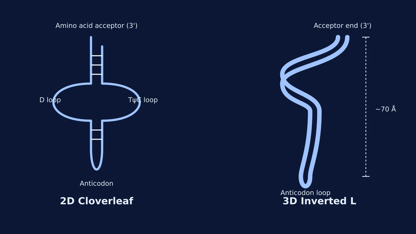
Transfer RNA acts as the cell's adapter molecule, reading genetic codes on mRNA and delivering the correct amino acid. While its flattened secondary form is the tRNA clover leaf structure, its actual three-dimensional shape is an inverted L, holding the anticodon loop and amino acid about 70 Å apart.
Before scientists mapped out the exact folds of nucleic acids, Francis Crick recognised a mechanical problem in the cell: amino acids cannot read the genetic code on their own. In 1958, he proposed an "adapter molecule" that could bridge the gap (source).
Today, we know this adapter as transfer RNA (tRNA). Previously called soluble RNA because of its small size, it does exactly what Crick predicted. It reads the instructions on messenger RNA (mRNA) and carries the correct amino acid to the growing protein chain. If you are learning the central dogma, such as three RNA polymerases in eukaryotes or RNA processing in eukaryotes: capping, tailing and splicing, understanding the structure of tRNA is a key step.
What is the adapter role of tRNA?
The adapter role means the tRNA molecule sits perfectly between two different biological languages. One end of the molecule interacts with the nucleic acid sequence of the mRNA, while the other end physically carries a specific amino acid.
Amino acids have no chemical affinity for the RNA sequence that codes for them. By acting as a physical bridge, this dual capability ensures that the genetic code translates accurately into a functional protein during translation.
Why does the tRNA clover leaf structure form?
Transfer RNA is a single strand of RNA. Because some of its bases are complementary to each other, the strand folds back on itself and forms base pairs through hydrogen bonding. When you flatten this secondary structure onto a page, it looks like a cloverleaf with three main loops and a stem.
Every tRNA has critical functional regions that carry out translation:
The amino acid acceptor end: This is the 3' end of the RNA strand, which always finishes with the sequence CCA. The specific amino acid attaches here during a process called aminoacylation.
The anticodon loop: Located at the bottom of the cloverleaf, this loop holds three exposed bases. These bases pair directly with the complementary codon on the mRNA.
The D loop: This region helps the specific aminoacyl-tRNA synthetase enzyme recognise the tRNA so it can attach the correct amino acid.
The TψC loop: This loop helps anchor the tRNA to the ribosome during protein synthesis.
While there are specific tRNAs for almost every codon, there are no tRNAs for the stop codons (UAA, UAG and UGA). When the ribosome reaches a stop codon, a release factor binds instead, terminating the chain.
How does the wobble hypothesis help the anticodon loop?
Francis Crick also noticed that while there are many different codons for amino acids, cells do not have a matching tRNA for every single one of them. He proposed the wobble hypothesis to explain this efficiency.
According to this rule, the first two bases of the mRNA codon pair strictly with the anticodon. However, the third base position can "wobble" or form non-standard base pairs. This flexibility allows a single tRNA to read multiple synonymous codons, ensuring translation continues smoothly without needing a massive variety of adapter molecules.
Why is the 3D structure an inverted L shape?
The cloverleaf is just a flattened two-dimensional map. In reality, the loops and stems fold further, twisting the molecule into a highly compact, three-dimensional form.
In 1974, Sung-Hou Kim and Alexander Rich published the first high-resolution structure of a yeast tRNA molecule using X-ray crystallography (source). They proved that the secondary cloverleaf folds into a distinct inverted L shape through complex base stacking and tertiary hydrogen bonds.
This shape is mechanically perfect for its adapter role. The acceptor stem and the TψC loop stack to form one long, rigid arm of the L, while the D loop and the anticodon loop form the other perpendicular arm. This arrangement places the anticodon at one extreme tip and the amino acid acceptor end at the opposite tip, about 70 Å apart.
This rigid distance allows the tRNA to plug its anticodon into the mRNA deep inside the decoding centre of the ribosome, while safely holding its amino acid precisely where the new peptide bond forms. If you are trying to picture how these structures fit together, the Fermi Concept Library has every concept in physics, chemistry, biology, mathematics and computing, each with its own lesson and its place in the map.
Know the ends: The 3' end is the amino acid acceptor end. It carries the CCA sequence.
Know the pairs: The anticodon on the tRNA pairs directly with the codon on the mRNA.
Know the structures: The clover leaf is the 2D secondary structure; the inverted L is the 3D tertiary structure.
Know the exceptions: Initiator tRNA starts the process, but there are no tRNAs for stop codons.


