

On this page
When Watson and Crick proposed the DNA double helix, they suggested that the molecule copies itself by splitting down the middle, with each half building a new complementary partner. While this elegant idea made sense for naked DNA, scientists needed to know if it also applied to the massive, protein-wrapped chromosomes of plants and animals.
In 1957, J. Herbert Taylor, Philip S. Woods and Walter L. Hughes proved that it does. Published in the *Proceedings of the National Academy of Sciences*, their experiment showed that entire eukaryotic chromosomes replicate semiconservatively, providing the first visual evidence of the process [[1]](https://doi.org/10.1073/pnas.43.1.122).
What did Taylor use for the experiment?
Taylor's team needed to see chromosomes clearly and track the new DNA as it was built. They chose three main tools:
***Vicia faba* (the broad bean):** The root tip cells of the faba bean divide rapidly and contain large, distinct chromosomes that are easy to view under a microscope.
Tritiated thymidine (³H-thymidine): Thymidine is a chemical building block used only to make DNA, never RNA. By tagging it with tritium (a radioactive isotope of hydrogen), the team ensured that only newly synthesised DNA would emit radiation.
Colchicine: This chemical stops cells from dividing by preventing the spindle apparatus from forming. It traps the cell at metaphase, keeping sister chromatids joined together at the centromere so their radiation can be compared side by side.
How did the Vicia faba experiment work?
The researchers tracked the bean chromosomes through two complete rounds of copying.
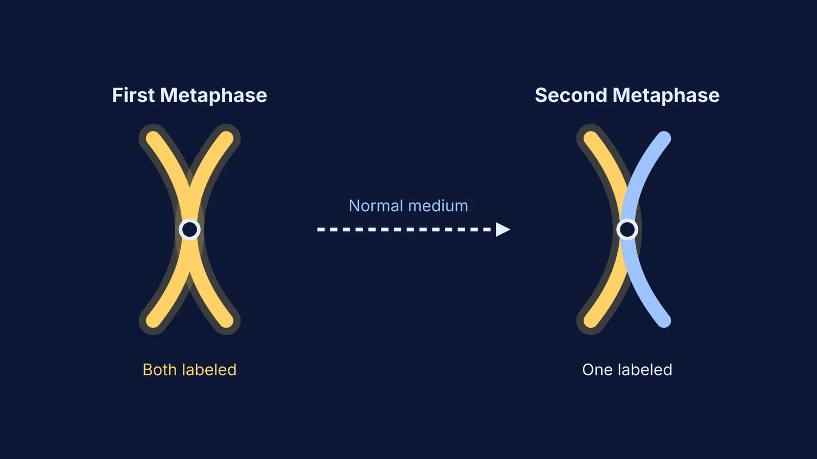
First, they grew the bean roots in a solution containing the radioactive thymidine. They allowed the cells to complete one cycle of DNA synthesis. When they examined the chromosomes at metaphase, both sister chromatids of every single chromosome were radioactive.
Next, they washed the roots and moved them to a normal, non-radioactive medium that contained colchicine. The cells duplicated their DNA a second time. When the researchers examined this second metaphase, the result was visually striking: in each chromosome, only one of the two sister chromatids was radioactive. The other chromatid was completely unlabeled.
What is autoradiography?
To actually see the radioactive DNA, the team used a technique called autoradiography.
When the radioactive tritium in the bean roots decayed, it released low-energy beta particles. The researchers placed a photographic emulsion over the squashed root cells. The beta particles hit the emulsion, turning the silver halides in the film into solid, dark grains of silver.
Under a microscope, these dark grains appeared precisely over the sections of the chromosome that contained the newly built DNA. Because tritium's beta particles travel only a very short distance before stopping, the image resolution was incredibly high—sharp enough for the team to tell exactly which of the two sister chromatids held the radioactive label.
Why did only one chromatid stay radioactive?
The findings perfectly matched the predictions of the semiconservative model.
Before the experiment began, the bean's DNA was entirely non-radioactive. During the first replication in the radioactive solution, the two original DNA strands separated. Each built a new, radioactive partner strand. Because each chromatid now contained one normal strand and one radioactive strand, both chromatids appeared labeled.
When the cells replicated again in the normal medium, those strands separated once more. The radioactive strand built a non-radioactive partner, creating a labeled chromatid. The non-radioactive strand also built a non-radioactive partner, creating a completely unlabeled chromatid.
If you find it hard to picture how DNA strands separate and rebuild during the cell cycle, the Fermi Concept Library offers a dedicated lesson for every concept in molecular biology, showing exactly where it fits in the wider map.
Taylor vs. Meselson and Stahl
This logic also underpins the Meselson and Stahl experiment, which proved the same mechanism. However, their approaches differed:
The subject: Taylor used a eukaryote (*Vicia faba*), showing that complex chromosomes follow the rule. Meselson and Stahl used a prokaryote (*E. coli*), which has simple, circular DNA.
The label: Taylor used a radioactive label (³H) to emit detectable particles. Meselson and Stahl used a heavy stable isotope (¹⁵N) to change the physical weight of the DNA.
The technique: Taylor looked at entire chromosomes using autoradiography. Meselson and Stahl extracted the DNA and spun it in a centrifuge.
Comparing these methods helps clarify how DNA replication and transcription differ across the domains of life.
A checklist for exam questions on classic experiments
When exams question you on classic biological experiments, they usually target specific design choices. Use this checklist to structure your revision:
The organism: Did they use *Vicia faba* (faba beans), *E. coli* or a bacteriophage?
The label: Was it a radioactive isotope (like ³H or ³²P) or a heavy isotope (like ¹⁵N)?
The technique: Did they use autoradiography to expose a film, or density gradient centrifugation to weigh the molecules?
The generation: Are you being asked about the first division (where everything is hybrid) or the second division (where the ratio changes)?
Working through the logic of these historical tests is far more effective than just memorising the result. Fermi's AI tutor can help you test your understanding by asking you to predict what Taylor would have seen if replication were conservative instead, checking your reasoning step by step.


