

On this page
Why does the iodine test produce a deep blue colour in starch?
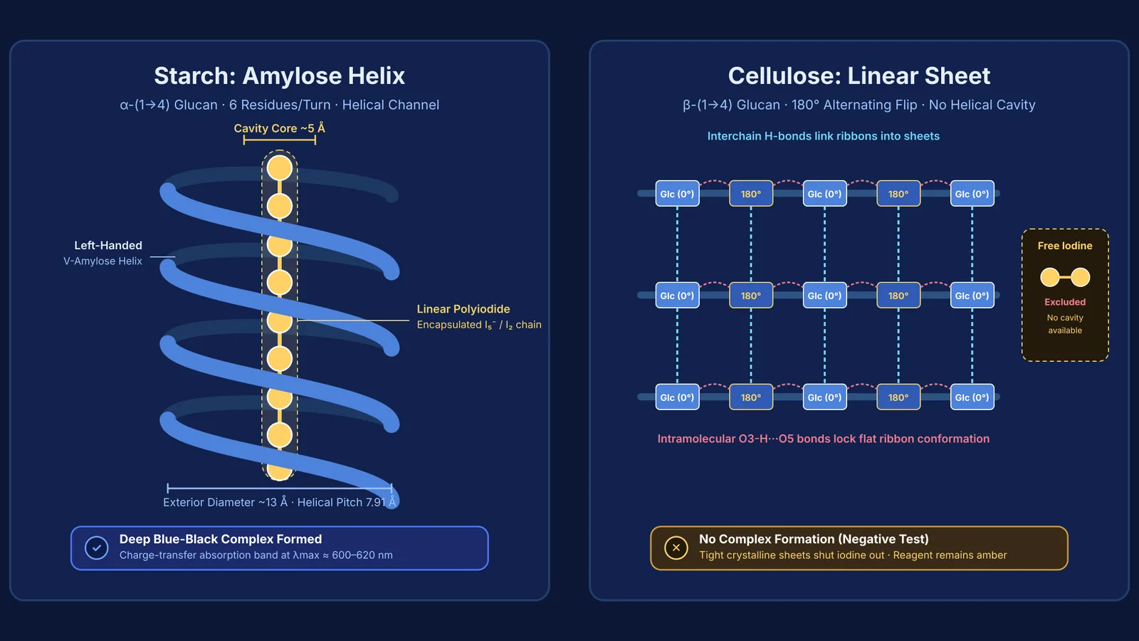
Adding a drop of yellowish-brown Lugol's iodine reagent to a starch suspension instantly produces an intense blue-black colour. This reaction depends on a supramolecular host-guest relationship between polyiodide ions and amylose, the unbranched helical fraction of plant starch. Amylose consists of D-glucose units joined by α-(1→4) glycosidic bonds, as explained in our guide to cyclic structures and anomers of glucose.
Because the α-(1→4) bond angles cause each pyranose ring to tilt relative to the next, amylose does not lie flat. Instead, it coils into a left-handed helix known as V-amylose. X-ray diffraction measurements by Robert E. Rundle and Dexter French (1943) showed that V-amylose contains six glucose residues per turn with a helical pitch of 7.91 Å and an exterior diameter of 13 Å. Its hydrophilic hydroxyl groups face outward toward water, leaving an internal hydrophobic core approximately 5 Å wide.
In potassium iodide solution, dissolved iodine forms linear polyiodide species. A 2024 review by Szilard Pesek and Radu Silaghi-Dumitrescu indicates that repeating I₂-I₅⁻-I₂ units slide into this central cavity. Within the helix, electron delocalisation between the polyiodide anions and neutral iodine produces a charge-transfer band with an absorption maximum near 600 nm to 620 nm. By absorbing orange-red light, the complex transmits a deep blue colour. You can see how this test works alongside others in our guide to laboratory tests for carbohydrates, fats and proteins.
How does the β-(1→4) linkage prevent cellulose from binding iodine?
Cellulose is also a homopolymer of D-glucose, yet it yields no colour change with iodine. This contrast stems entirely from glycosidic stereochemistry: cellulose is linked by β-(1→4) bonds rather than α-(1→4) bonds.
To connect equatorial hydroxyl groups at carbons 1 and 4 without ring strain, every alternating glucose unit is flipped 180°. Synchrotron X-ray and neutron diffraction by Yoshiharu Nishiyama, Paul Langan, and Henri Chanzy (2002) demonstrated that this flip prevents coiling, locking cellulose into flat, extended ribbons.
Intramolecular O3-H···O5 hydrogen bonds stiffen each chain, while interchain hydrogen bonds bind adjacent ribbons into crystalline sheets. Because these sheets pack tightly into microfibrils, cellulose contains no cylindrical cavities or hydrophobic tunnels. Free iodine cannot enter, leaving the solution a faint amber.
Why does heating make the blue starch-iodine complex disappear?
Heating the blue mixture causes the colour to vanish, leaving a clear or pale yellowish liquid. When the solution cools back to room temperature, the deep blue colour returns.
This reversible bleaching demonstrates that the complex relies on non-covalent confinement rather than covalent bonds. Thermal energy disrupts the hydrogen bonds and hydrophobic interactions that hold the amylose helix in shape, unravelling the polymer into a flexible random coil. Without a rigid cylindrical host, the polyiodide chain dissociates into individual iodide and triiodide species that do not absorb visible light in the orange-red spectrum. Upon cooling, the amylose chains re-coil into helices and re-encapsulate the polyiodides.
For molecular shapes that are difficult to picture from flat textbook drawings, the Fermi Simulation Lab provides interactive simulations for ideas that are easier to see than to read.
How do competitive exams test polysaccharide conformation?
Examiners in CBSE and NEET-UG regularly test whether students understand how bond stereochemistry dictates macromolecular shape. The NCERT Class 11 Biology curriculum explicitly notes that starch forms helical secondary structures that hold iodine, whereas cellulose lacks complex helices and cannot hold iodine. A four-step diagnostic framework clarifies these questions:
Identify linkage geometry: α-(1→4) bonds curve into helices, while β-(1→4) bonds create flat, extended ribbons.
Evaluate cavity formation: an open helical channel accommodates polyiodides, whereas dense interchain packing in cellulose sheets excludes them.
Check branching effects: extensive branching in amylopectin restricts continuous helical stretches, yielding a reddish-purple hue rather than deep blue.
Assess thermal reversibility: helical inclusion complexes decolourise upon heating as coils unravel, while cellulose remains unreactive at all temperatures.
You can trace how these principles apply to other macromolecules in our guide to biomolecules in chemistry.
To see how glycosidic bond stereochemistry connects to broader carbohydrate classification, the Fermi Concept Library covers every concept in physics, chemistry, biology, mathematics and computing, each with its own lesson and its place in the map.


