

On this page
Why does pollen survive for hundreds of millions of years?
When botanists examine sedimentary rocks from deep geological time, delicate leaves, flowers, and stems have almost always vanished. Yet pollen grains and plant spores appear under the microscope with their intricate walls, spikes, and geometric ridges intact. Charles H. Wellman and colleagues reported in Nature that fossilised cryptospores from Oman, dated to approximately 475 million years ago during the Middle Ordovician, survive because their outer coat contains sporopollenin—the most resilient biopolymer synthesized by living organisms.
For nearly two centuries, sporopollenin resisted chemical characterisation because standard laboratory reagents could not dissolve it. In 2019, a research team led by Jing-Ke Weng at MIT solved this problem using thioacidolysis and solid-state nuclear magnetic resonance spectroscopy. As published in Nature Plants, pine sporopollenin is a complex heteropolymer built of polyvinyl alcohol units and 7-O-p-coumaroylated C16 aliphatic chains crosslinked through m-dioxane acetal rings, alongside aromatic phenolics like p-coumaric acid and naringenin.
This dense crosslinking protects the male gametophyte from temperatures up to 250 °C, concentrated mineral acids, strong alkalis, and microbial decay. In palynology, researchers isolate pollen walls using Gunnar Erdtman's classic acetolysis procedure, reviewed in Plant Systematics and Evolution. Boiling pollen in a 9:1 mixture of acetic anhydride and concentrated sulphuric acid dissolves all internal cytoplasm, intine, and nucleic acids, leaving only the unaltered sporopollenin skeleton.
How do the exine and intine wall layers differ?
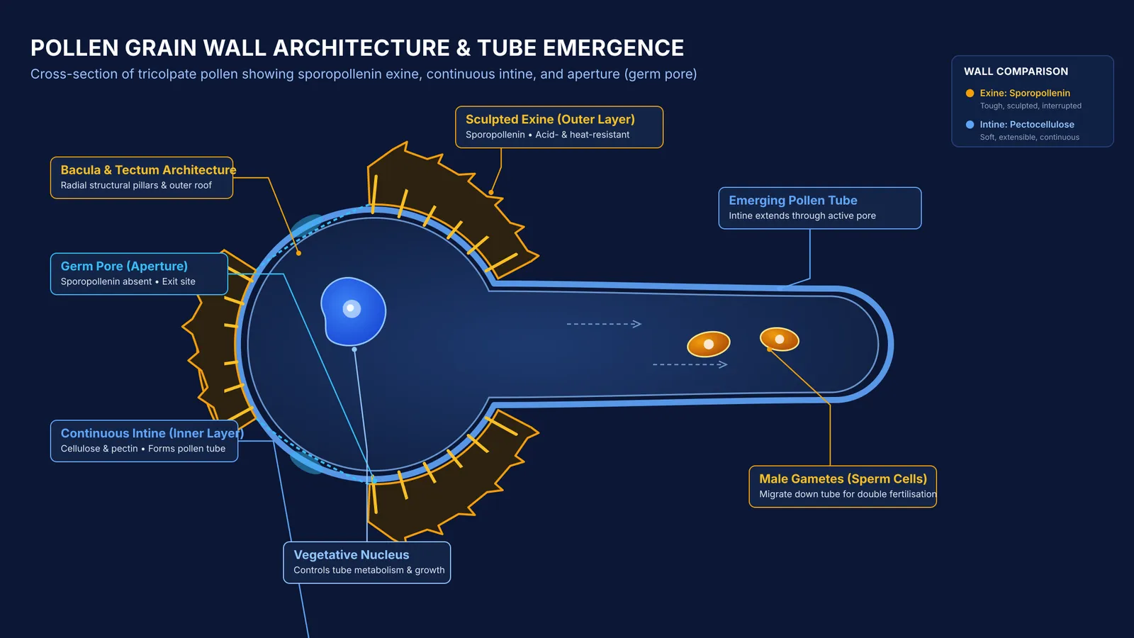
The pollen grain wall, known scientifically as the sporoderm, consists of two distinct layers with complementary roles:
The exine: The tough, sculpted outer layer made of sporopollenin. It displays species-specific ridges, reticulations, or spines. As reviewed in the Annual Review of Plant Biology, the tapetum—the nutritive inner layer of the anther—deposits sporopollenin precursors onto developing grains during microsporogenesis and pollen grain development. Mature pollen grains typically measure between 25 and 50 micrometres in diameter.
The intine: The soft, flexible inner layer beneath the exine. It contains no sporopollenin; instead, it consists of cellulose, hemicellulose, and pectin. Synthesized directly by the pollen protoplast, the intine forms a thin, continuous boundary around the vegetative cell.
While the exine shields gametes against ultraviolet radiation and mechanical damage during dispersal, the intine provides the extensible matrix required for tube growth. Successful pollination later unites these gametes with the female gametophyte formed during megasporogenesis and embryo sac formation.
Why must germ pores lack sporopollenin?
Because no known plant, microbial, or animal enzyme can digest sporopollenin, an unbroken exine shell would permanently trap the male gametes inside. To permit germination, the exine leaves deliberate openings called germ pores (or apertures).
In The Plant Cell, Anna A. Dobritsa and Daniel Coerper demonstrated that aperture formation is genetically controlled. The protein INAPERTURATE POLLEN1 (INP1) localises to specific plasma membrane bands during microsporogenesis, preventing sporopollenin deposition at those sites. Without functional INP1, microspores deposit sporopollenin uniformly across their entire surface, producing completely apertureless pollen grains.
Apertures also provide mechanical flexibility. In the Proceedings of the National Academy of Sciences, Anže Božič and Antonio Šiber showed that apertures are 15 to 100 times softer than the surrounding sporopollenin wall. In dry air, these soft regions fold inward—a reversible process called harmomegathy—sealing the grain against dehydration.
When pollen lands on a compatible stigma, hydration raises internal turgor pressure. A pectin-rich lens beneath the pore (the oncus) softens, allowing the intine to bulge outward through the aperture as the pollen tube. Agricultural breeders monitor this process closely during artificial hybridisation and emasculation.
How do you solve pollen wall questions in exams?
Questions on pollen wall architecture appear regularly in Class 12 board examinations and NEET-UG. Use this four-step checklist to avoid common traps:
Identify the layer: Exine is the tough outer sporopollenin wall; intine is the inner pectocellulosic wall.
Verify continuity: Intine is continuous around the protoplast; exine is discontinuous because germ pores interrupt it.
Trace the emerging tube: The pollen tube is an extension of the inner intine emerging through a germ pore.
Count the apertures: Monocots typically produce monosulcate pollen with one germ furrow, while dicots typically produce tricolpate pollen with three germ pores.
When revising plant reproduction, connecting microscopic features to developmental physiology helps prevent confusion in assertion-reason questions. The Fermi Concept Library organizes every concept in physics, chemistry, biology, mathematics and computing within a structured topic map. To build precision on exam-style questions, the Fermi Problem Library provides worked problems with a step-by-step solver that explains the reasoning rather than printing an answer.


