

On this page
The production of sperm requires a tightly controlled environment, managed largely by two specialised types of somatic cells in the testis: Sertoli cells and Leydig cells. Though they work toward the same ultimate goal of fertility, they sit in different microscopic compartments and perform very different physiological tasks.
Where do Sertoli and Leydig cells sit?
Testicular tissue is divided into two distinct compartments. The seminiferous tubules are the highly coiled, tube-like structures where germ cells divide and mature into sperm.
Sertoli cells sit inside the seminiferous tubules. They are large, columnar cells that stretch from the outer basement membrane of the tubule all the way to its central lumen. By locking together with tight junctions, they form the blood-testis barrier. This physical wall is essential because developing sperm cells are genetically distinct from the rest of the body; without the barrier, the immune system would mistakenly attack them as foreign invaders.
Leydig cells are interstitial cells, meaning they sit outside the tubules in the loose connective tissue spaces between them. They cluster near the blood vessels that supply the testis.
What is the function of Sertoli cells?
Often called "nurse cells," Sertoli cells are responsible for nourishing and protecting germ cells as they develop into mature sperm. Their activity is primarily stimulated by Follicle-Stimulating Hormone (FSH) secreted by the anterior pituitary gland.
Every step of spermatogenesis depends on this support network. Because a single Sertoli cell can only physically and metabolically sustain a limited number of developing sperm, the total Sertoli cell count acts as the primary biological bottleneck for sperm production capacity. When you study how these specific cells fit into the broader picture of the cell as the unit of life, their structural adaptations for secretion and support become clear. To explore how FSH and other signals trigger cellular responses, the Fermi Concept Library has its own lesson on the mechanics of endocrine signaling and its place in the map.
What is the function of Leydig cells?
Leydig cells are the steroid-producing factories of the testis. Stimulated by Luteinizing Hormone (LH) from the pituitary gland, their main function is to synthesize and secrete the androgen testosterone.
While some of this testosterone enters the general bloodstream to drive secondary male characteristics, a massive amount of it must be kept locally in the testis to support sperm development. Sertoli cells produce androgen-binding protein (ABP) to trap this testosterone where it is needed. In healthy men, intratesticular testosterone levels are approximately 40 times higher than the levels found in peripheral blood (Coviello et al., 2004). If this local concentration drops—even if blood testosterone levels remain seemingly normal—sperm production halts entirely.
How many of these cells are in a human testis?
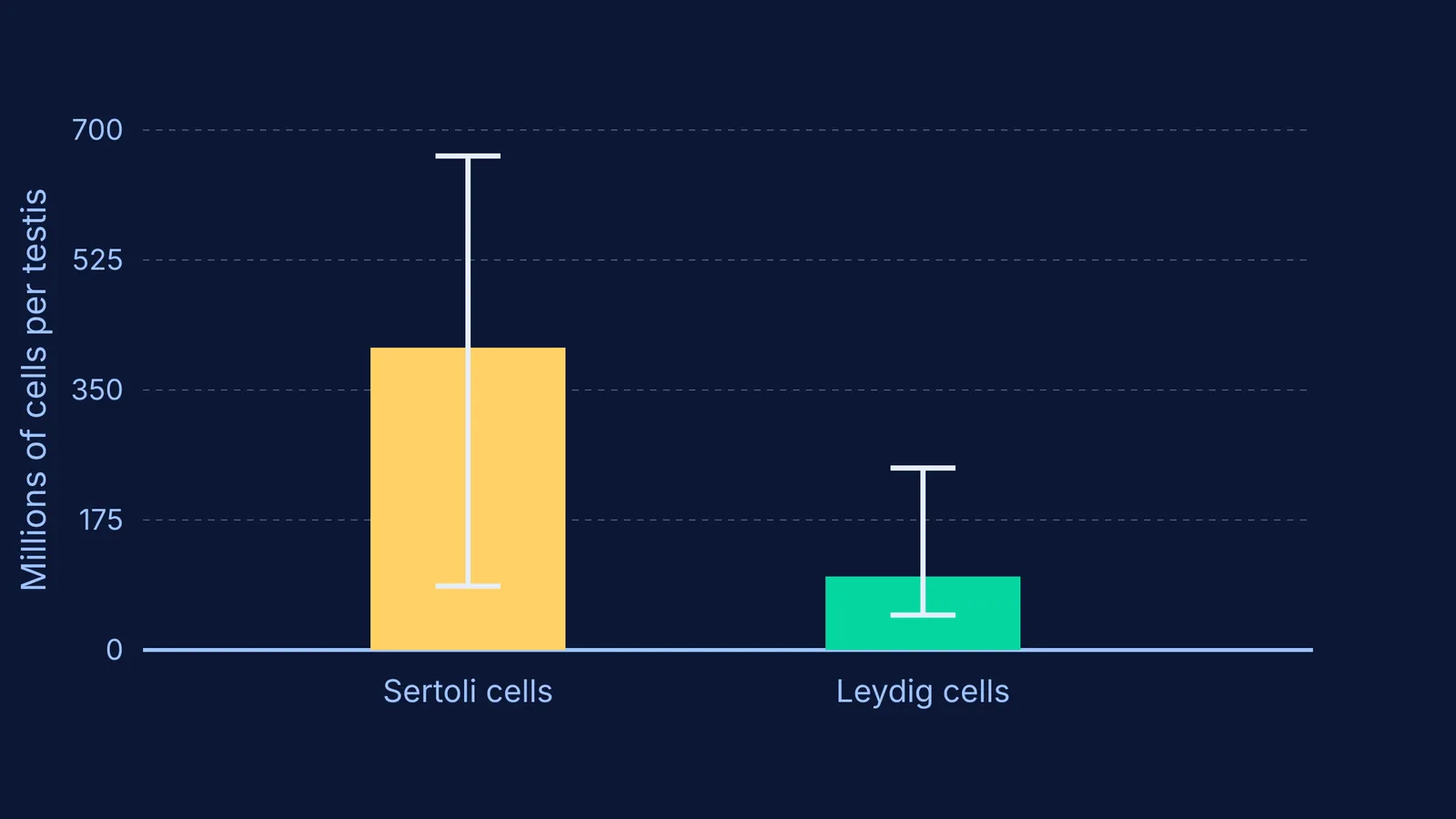
Because these cells are so critical to fertility, researchers have meticulously quantified them. A landmark stereological study found that a single adult human testis contains a mean of 407 million Sertoli cells and 99 million Leydig cells (Petersen et al., 2015).
The researchers noted that while the total number of Leydig cells remains relatively stable, the population of Sertoli cells declines significantly as men age. Because Sertoli cell numbers dictate the maximum possible sperm output, this progressive loss is a key factor in changing male fertility over time.
A practical framework for exam prep
These two cell types are heavily tested in biology curricula and NEET-UG. When mapping out hormonal feedback loops or studying the clinical aspects of reproductive health, you can quickly distinguish them using a simple three-point checklist:
Location: Sertoli cells are strictly inside the tubules; Leydig cells are in the interstitial space outside.
Hormone trigger: Sertoli cells are driven by FSH; Leydig cells are driven by LH.
Primary output: Sertoli cells provide physical nourishment and form barriers; Leydig cells secrete testosterone.


