

On this page
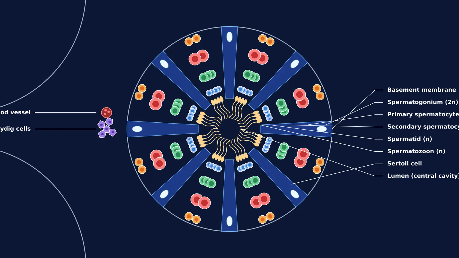
Inside the human testes, sperm are manufactured in highly coiled tubes called seminiferous tubules. If you cut across one of these tubes and look under a microscope, you see a highly organised factory floor. This transverse section (T.S.) is a classic diagram for board exams and NEET biology, as it maps the exact sequence of male germ cell development.
What are the layers of a seminiferous tubule from edge to centre?
Spermatogenesis is a one-way journey from the tubule's outer wall (the basement membrane) to its hollow centre (the lumen). The entire developmental process takes approximately 64 days in humans (Heller & Clermont, 1963). As cells divide and mature, they are pushed progressively closer to the centre.
Spermatogonia: These are the diploid stem cells sitting directly on the basement membrane. They divide continuously by mitosis. Some of the new cells remain as stem cells, while others are committed to becoming sperm.
Primary and secondary spermatocytes: The committed spermatogonia grow and move inward to become primary spermatocytes. These large cells undergo the first meiotic division to form haploid secondary spermatocytes. (This reduction in chromosome number is mirrored in plants; see Microsporogenesis and the structure of a pollen grain).
Spermatids: The secondary spermatocytes rapidly undergo the second meiotic division. The result is spermatids—small, round, immature cells that finally contain the correct haploid genome but have no tails.
Spermatozoa (Sperms): In the final phase, called spermiogenesis, spermatids shed excess cytoplasm and grow flagella (tails) to become mature, motile spermatozoa. Their heads remain embedded in nurse cells for nourishment, while their tails point directly into the central lumen, ready to be washed out of the testis.
What is the role of Sertoli and Leydig cells?
The developing germ cells do not work alone. Two other vital cell types complete the diagram:
Sertoli cells: These tall, pillar-like cells stretch entirely from the basement membrane to the lumen. Often called nurse cells, they provide structural support and nourishment to the developing sperm. Crucially, tight junctions between adjacent Sertoli cells form the blood-testis barrier. Because haploid sperm cells have a different genetic makeup from the rest of the body, this barrier hides them from the immune system, preventing an autoimmune attack.
Leydig cells (Interstitial cells): These are found entirely *outside* the tubule, occupying the connective tissue spaces between adjacent tubules. Under the influence of the pituitary gland, Leydig cells secrete androgens, primarily testosterone. This hormone diffuses into the tubules to drive the entire sperm-production process. To see how hormones like testosterone trigger responses, you can read How hormones act: membrane receptors vs intracellular receptors.
How long does spermatogenesis take?
While it takes approximately 64 days for a single spermatogonium to become a mature spermatozoon, the male body produces sperm continuously every day. This happens because the tubule operates in overlapping cycles. A new cycle of the seminiferous epithelium begins approximately every 16 days (Heller & Clermont, 1963). At any given time, different patches along the length of a tubule are at different stages, ensuring a constant, continuous release of sperm into the lumen. Once released, the sperm spend an additional time maturing in the epididymis before they are fully capable of fertilisation (for the next steps in reproduction, see From fertilisation to implantation: the first week of pregnancy).
How can Fermi's AI tutor help with biology diagrams?
When you study complex structures like the seminiferous tubule, passive reading rarely makes it stick. If you get stuck on the sequence of meiotic divisions or keep confusing spermiogenesis with spermatogenesis, Fermi's AI tutor can work out what you already know and build a plan for the session. By checking your understanding as it goes, Fermi ensures you know the biological reasons behind the diagram's layout, rather than just memorising the shapes. For a deep dive into male reproductive anatomy and thousands of other topics, you can explore the Fermi Concept Library.
How do you draw the T.S. of a seminiferous tubule?
If you need to reproduce this diagram in an exam, use this mental checklist to ensure you do not drop marks:
The boundary: Draw a large, neat circle for the basement membrane.
The support: Add a few tall, wavy triangles reaching from the edge to the centre for the Sertoli cells.
The gradient: Draw the germ cells in distinct layers. Put cuboidal spermatogonia at the edge, larger primary spermatocytes in the middle, and small round spermatids near the centre.
The tails: Draw spermatozoa with wavy tails pointing directly into the central cavity (lumen), not sideways or outwards.
The exterior: Draw a cluster of polygonal Leydig cells with a few blood vessels entirely outside the main circle.


