

On this page
What is a satellite chromosome in cell biology?
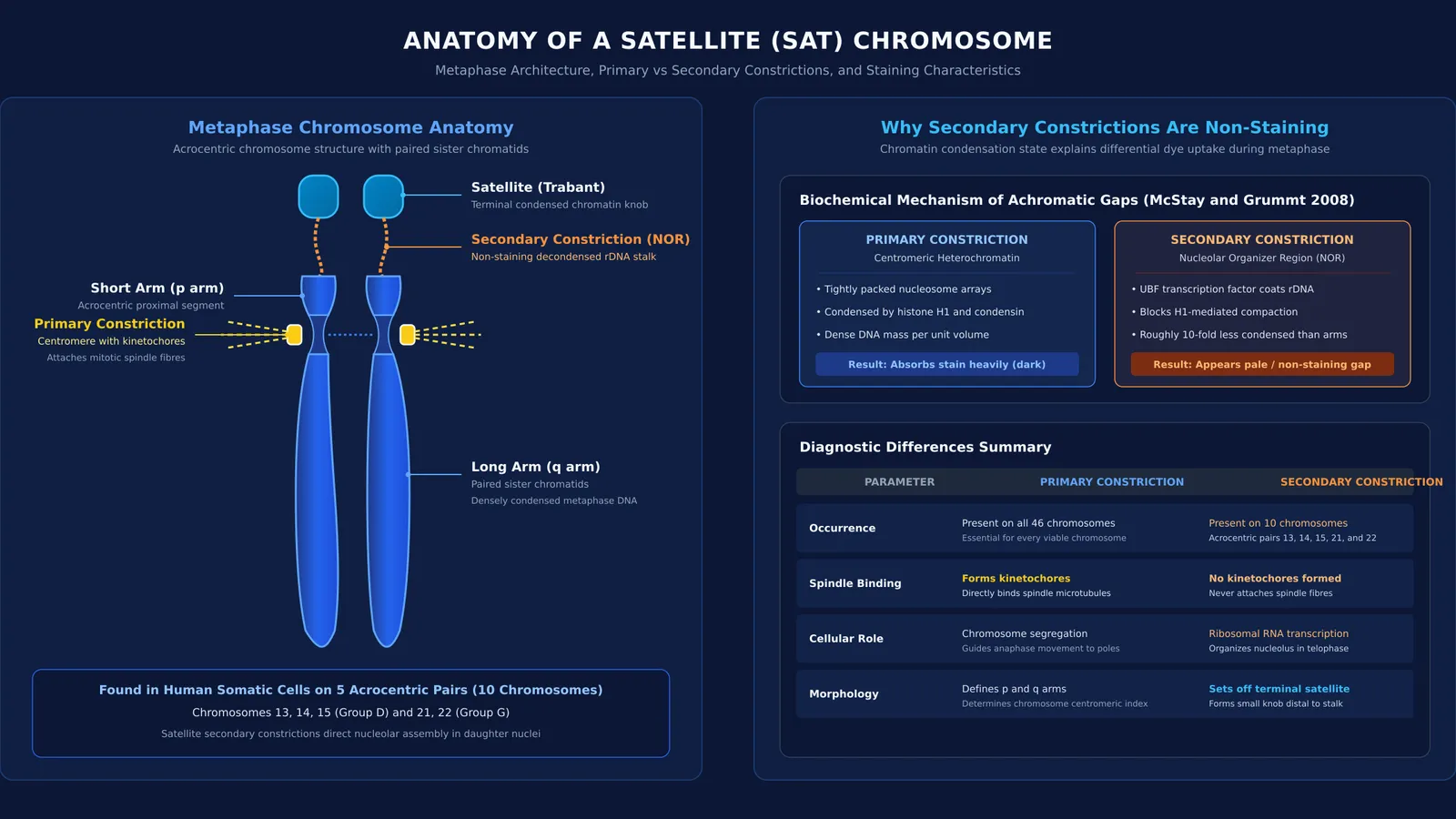
When eukaryotic cells enter mitotic metaphase, every chromosome exhibits a primary constriction called the centromere. This narrow waist holds sister chromatids together and organizes the kinetochores that bind spindle fibres. However, certain chromosomes feature an additional morphological indentation called a secondary constriction. A satellite chromosome is any chromosome that possesses a non-staining secondary constriction at a constant site, setting off a small terminal chromatin fragment called the satellite.
Distal to this constriction, the satellite appears as an isolated knob under a light microscope, connected to the chromosome arm by a slender chromatin stalk. As explored in our guide to types of chromosomes based on centromere position, chromosome shape during anaphase depends on the centromere, but the presence and position of a satellite remain constant for a given chromosome pair.
Why does NCERT call secondary constrictions non-staining?
The NCERT Class 11 Biology syllabus highlights that "sometimes a few chromosomes have non-staining secondary constrictions at a constant location." This phrase often puzzles students. When cytogeneticists prepare chromosome spreads, they apply basic stains such as acetocarmine, Giemsa, or Feulgen reagent. These dyes bind intensely to condensed chromatin, turning the chromosome arms dark purple. Secondary constrictions appear pale under brightfield microscopy not because they lack DNA, but because their chromatin remains undercondensed.
In a foundational study in Planta, Emil Heitz (1931) showed that nucleoli form at specific chromosomal constrictions, coining the term "SAT chromosome" from the Latin phrase *sine acido thymonucleinico* (without thymonucleic acid) because the pale constriction failed to show standard Feulgen staining for DNA. Subsequently, Barbara McClintock (1934) proved in maize that this secondary constriction corresponds directly to the nucleolar organizer region.
Modern molecular cytogenetics explains why this locus resists condensation. As reviewed by Brian McStay and Ingrid Grummt in the Annual Review of Cell and Developmental Biology, nucleolar organizer regions contain tandem ribosomal DNA repeats. The transcription factor UBF binds across active ribosomal DNA throughout cell division. This binding prevents histone H1 from condensing the chromatin, leaving the active locus roughly 10-fold less condensed than the surrounding metaphase arms. Because DNA concentration per unit volume in this uncoiled stalk is much lower, it absorbs negligible stain and appears as a non-staining gap. For students mastering cytology fundamentals, the Fermi Concept Library covers every concept in physics, chemistry, biology, mathematics and computing, each with its own lesson and its place in the map.
Which human chromosomes have secondary constrictions and satellites?
In human somatic cells, satellites occur exclusively on the short arms of the five acrocentric autosomes: chromosomes 13, 14, 15, 21, and 22. In a classic radio-labeling study in the Proceedings of the National Academy of Sciences, A. S. Henderson, D. Warburton, and K. C. Atwood confirmed that ribosomal DNA localizes specifically to these five chromosome pairs. Because human somatic cells are diploid with 46 chromosomes, exactly 10 chromosomes carry these secondary constrictions and satellites.
Modern sequencing by Marjolein van Sluis and colleagues in Genes & Development confirmed that these five chromosome pairs share conserved distal junctions coordinating nucleolar assembly. Understanding this architecture—alongside our Class 11 cell structure and organelle overview—clarifies how multiple chromosomes join to organize the nucleolus.
How do primary and secondary constrictions differ?
To avoid exam traps, compare primary and secondary constrictions across these parameters:
Occurrence: The primary constriction (centromere) is present on all normal chromosomes, whereas secondary constrictions occur only at constant sites on specific chromosomes.
Spindle binding: The primary constriction forms kinetochores that attach spindle fibres, while secondary constrictions never bind spindle fibres.
Staining: The primary constriction stains darkly due to condensed heterochromatin, whereas the secondary constriction appears non-staining because of undercondensed ribosomal DNA.
Function: The primary constriction directs chromosome segregation, whereas the secondary constriction transcribes ribosomal RNA to organize the nucleolus.
Satellite: The primary constriction defines p and q arm lengths, whereas the secondary constriction sets off the terminal satellite knob.
Understanding how chromatin compacts—as outlined in our guide to eukaryotic chromatin packaging and nucleosomes—clarifies why ribosomal loci remain decondensed.
How should you tackle satellite chromosome questions in NEET?
NEET questions on chromosome morphology frequently test structural definitions. Use this 3-step checklist:
Check the spindle fiber trap: Spindle fibres bind exclusively to kinetochores at the primary constriction. Any option claiming they attach to secondary constrictions is false.
Count chromosomes accurately: As demonstrated by Henderson et al. (1972), humans have 5 acrocentric satellite pairs. A diploid somatic cell has 10 satellite chromosomes, while a haploid gamete has 5.
Distinguish SAT chromosomes from satellite DNA: Satellite DNA consists of highly repetitive non-coding sequences separated by density centrifugation, whereas a SAT chromosome is a physical chromosome bearing a secondary constriction and satellite.
When working through multi-statement biology problems, the Fermi Problem Library provides worked problems with a step-by-step solver that explains the reasoning rather than printing an answer. If you want to identify specific conceptual gaps in cell division or genetics, Fermi's AI tutor can work out what you already know and build a plan for the session.


