

On this page
Every minute of the day, your body adjusts the depth and rate of your breathing to match your metabolic needs. This automatic process is controlled by a specialised neural network in the brainstem. For students preparing for medical exams, understanding the exact roles of the respiratory rhythm centre and pneumotaxic centre is a key part of the biology syllabus.
Where is the respiratory rhythm centre located?
The main pacemakers for your breathing sit in the medulla oblongata, the lower half of the brainstem. The respiratory rhythm centre is primarily responsible for generating and maintaining the basic rhythm of breathing (Source: Wikipedia). It contains clusters of neurons that rhythmically fire impulses down the phrenic nerve, instructing the diaphragm and external intercostal muscles to contract for inspiration.
When you are resting, this centre ensures a steady, quiet breathing pattern, known as eupnea. It acts much like a metronome, initiating each breath without any conscious thought on your part. If you need more air—such as during strenuous exercise—the rhythm centre can recruit accessory muscles in the chest and abdomen to force deeper, more rapid exhalations, clearing the lungs completely before the next breath.
What is the function of the pneumotaxic centre?
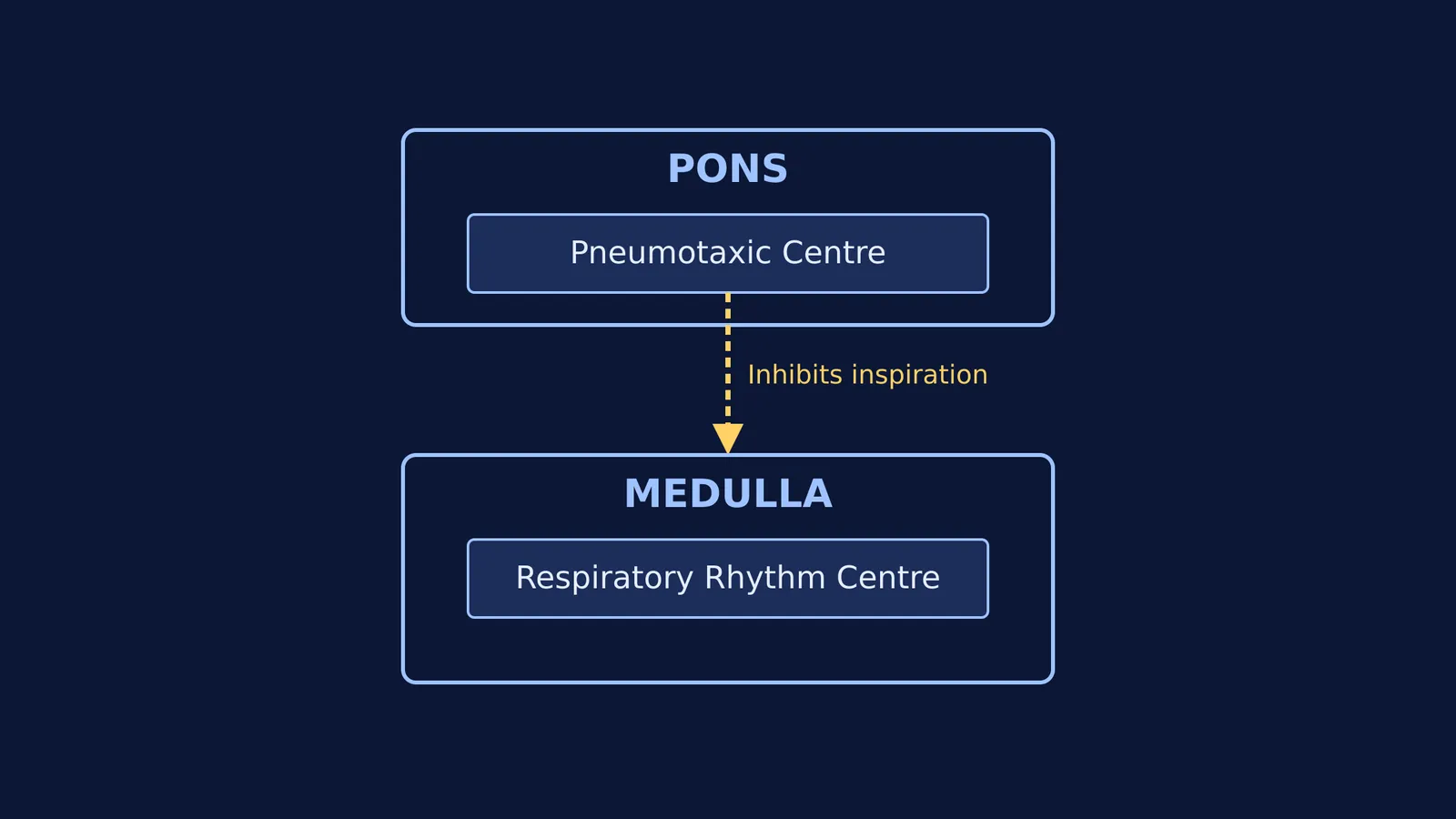
While the medulla generates the rhythm, it needs continuous moderation to adapt to what you are doing. This is where the pons, located just above the medulla, comes in. The pneumotaxic centre in the pons acts as an "off-switch" for the inspiratory signal (Source: Wikipedia).
By sending inhibitory neural signals down to the respiratory rhythm centre, the pneumotaxic centre can actively reduce the duration of inspiration. When each breath is cut slightly shorter, the respiratory rate automatically increases to compensate. This vital mechanism helps prevent the lungs from over-inflating and allows the body to transition smoothly and quickly from inhalation to exhalation.
Why is the role of oxygen in respiratory rhythm insignificant?
One of the most frequent traps in exams is assuming that a sudden drop in oxygen drives our normal urge to breathe. In reality, under most conditions, the partial pressure of carbon dioxide in the bloodstream is the most important factor controlling the respiratory rate, rather than oxygen levels (Source: Wikipedia). The role of oxygen in the normal day-to-day regulation of respiratory rhythm is quite insignificant.
Instead, the primary drivers are carbon dioxide (CO₂) and hydrogen ions (H⁺). Because CO₂ dissolves in blood to form carbonic acid, which releases H⁺, the two are closely linked. The brain detects these changes through two main systems:
Central chemoreceptors: Situated right next to the respiratory rhythm centre in the medulla, this region is highly sensitive to CO₂ and H⁺. When it detects a rise in these chemicals, it activates the rhythm centre to immediately increase the breathing rate, venting off the excess CO₂ through the lungs.
Peripheral chemoreceptors: Chemoreceptors associated with the aortic arch and carotid artery also recognise changes in CO₂ and H⁺ concentration. They send necessary signals up to the rhythm centre for remedial actions.
Only in extreme situations, such as severe hypoxia, does a lack of oxygen become a major regulatory factor.
How can you study this efficiently?
Because this is a dense topic, rote memorisation often fails under exam pressure. To study the respiratory rhythm centre effectively, try these practical steps:
Draw the pathway: Instead of just reading the text, draw a simple block diagram on a blank page. Place the pons above the medulla. Draw an arrow from the pneumotaxic centre down to the medulla, and label it "inhibits inspiration." The physical act of drawing helps lock the spatial relationship in your memory.
Group the receptors: Create a two-column table for the sensors. Put the central chemoreceptors in one column (noting they are central, located in the medulla) and the aortic and carotid chemoreceptors in the other (noting they are peripheral, located near the heart and neck). Note that both detect CO₂ and H⁺.
Write out the "oxygen trap": Write a large note to yourself that oxygen is not the primary driver. Remind yourself that a person holding their breath is forced to exhale not because they run out of oxygen, but because CO₂ builds up in the blood and severely triggers the central chemoreceptors.
Mastering biology concepts
Understanding complex regulatory loops requires seeing how all the pieces connect. It is much like tracing the metabolic steps of respiration as an amphibolic pathway or mapping out the feedback mechanisms of parturition and lactation.
When you encounter a mechanism that is hard to picture from a textbook paragraph alone, the Fermi Concept Library can help you break it down into manageable ideas. And if you find yourself confused by a tough exam question, Fermi's AI tutor works out what you already know and builds a plan for the session, so you sit down to a clear next step instead of staring at a blank page.


