

On this page
Which monomers and functional groups do these three bonds connect?
Biological macromolecules rely on covalent linkages to join building blocks into functional polymers. Living cells assemble proteins, carbohydrates, and nucleic acids using three distinct linkages: peptide bonds, glycosidic bonds, and phosphodiester bonds.
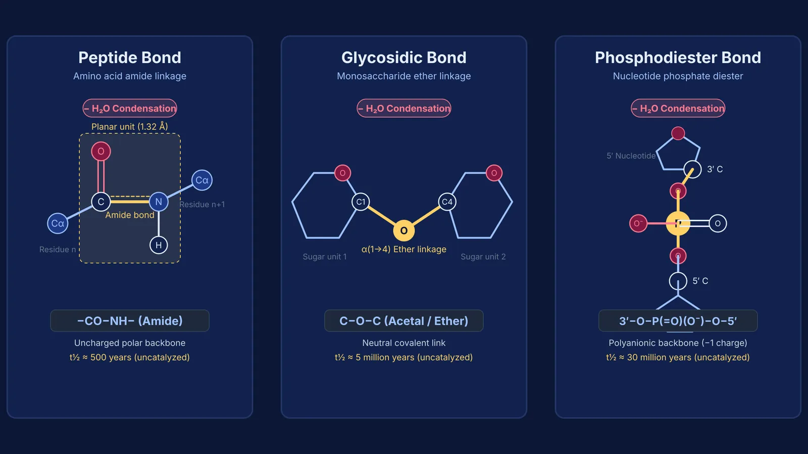
Peptide bonds link alpha-amino acids into chains. The alpha-carboxyl group (−COOH) of one amino acid reacts with the alpha-amino group (−NH₂) of the next to form an amide linkage (−CO−NH−). In Section 3.2 of *Biochemistry*, Jeremy M. Berg, John L. Tymoczko, and Lubert Stryer note that this planar bond has a carbon-nitrogen distance of 1.32 Å, midway between a single bond (1.49 Å) and a double bond (1.27 Å) (Berg et al., Section 3.2). Explore dipeptide naming in our guide to peptide bond formation and dipeptide nomenclature.
Glycosidic bonds join monosaccharides into carbohydrates. The anomeric carbon of one sugar—such as C1 in glucose—reacts with a hydroxyl group (−OH) on another sugar, forming an ether linkage (C−O−C) known as an acetal bridge (Berg et al., Section 11.2). Maltose links two glucose units via an alpha-1,4-glycosidic bond, while sucrose joins glucose and fructose via an alpha-1,2 bond, explaining why sucrose acts as a non-reducing sugar.
Phosphodiester bonds link nucleotides in DNA and RNA. A phosphate group bridges the 3′-hydroxyl of one pentose to the 5′-hydroxyl of the adjacent sugar (Berg et al., Section 5.1). Because phosphoric acid forms two ester bonds—one to each sugar—it creates a phosphodiester bridge. For monomer chemistry, see our guide on nucleosides and nucleotides in DNA and RNA.
Where does the water molecule come from during bond formation?
All three linkages form through condensation (dehydration synthesis), releasing one water molecule (H₂O) per bond formed:
In peptide bonds, −OH leaves the alpha-carboxyl group of the first amino acid, and −H leaves the alpha-amino group of the second.
In glycosidic bonds, −OH leaves the anomeric carbon of the first sugar, and −H leaves a hydroxyl group on the second.
In phosphodiester bonds, −OH leaves the phosphate moiety, and −H leaves the pentose 3′-hydroxyl.
Breaking these bonds requires hydrolysis, adding water across the linkage. Without enzymes, these linkages are exceptionally stable. Anna Radzicka and Richard Wolfenden reported in the *Journal of the American Chemical Society* that uncatalyzed peptide bond hydrolysis at 25 °C has a half-life of roughly 500 years (Radzicka and Wolfenden, 1996). For unactivated glycosidic bonds, Richard Wolfenden, Xiangdong Lu, and Gregory Young measured an uncatalyzed half-life of roughly 5 million years at 25 °C in model glycosides (Wolfenden et al., 1998). Because negative phosphate charges repel nucleophiles, Gottfried K. Schroeder and colleagues showed in the *Proceedings of the National Academy of Sciences* that a phosphodiester model has an uncatalyzed half-life of roughly 30 million years at 25 °C, accelerated 10¹⁷-fold by nucleases (Schroeder et al., 2006).
How do bond structures shape proteins, polysaccharides and nucleic acids?
The geometry and flexibility of each linkage govern macromolecular architecture:
Peptide bonds enforce planarity. Resonance restricts rotation around the C−N bond, limiting conformational flexibility to rotation around the alpha-carbon single bonds and guiding chains into alpha-helices and beta-sheets.
Glycosidic bonds determine polysaccharide shape. In starch and glycogen, alpha-1,4-linkages produce open helical coils that enzymes readily digest. Glycogen branches via alpha-1,6-bonds every 10 glucose units, while amylopectin in starch branches every 30 units (Berg et al., Section 11.2). Conversely, beta-1,4-linkages in cellulose form straight ribbons that pack into rigid microfibrils with high tensile strength.
Phosphodiester bonds provide polarity and stability. The 5′-to-3′ phosphoester backbone gives nucleic acids directionality. Each phosphate carries a negative charge at physiological pH, repelling spontaneous hydrolysis and stabilizing the double helix exterior.
When connecting structural chemistry to biological function, seeing how geometric constraints define molecular properties helps students build intuition. The Fermi Concept Library includes every concept in physics, chemistry, biology, mathematics and computing, each with its own lesson and its place in the map.
How can you calculate water loss and identify unknown bonds?
Exam problems frequently test these linkages through water loss stoichiometry or unknown bond identification, as detailed in standard structural biochemisty frameworks (Berg et al., Section 3.2).
Use this checklist to solve accounting problems and identify linkages:
Calculate water loss: An unbranched linear chain of n monomers forms n − 1 bonds and releases n − 1 water molecules. For instance, a chain of 100 amino acids has 99 peptide bonds and releases 99 water molecules. A single DNA strand of 100 nucleotides contains 99 phosphodiester bonds. In a closed cyclic polymer, the count equals n.
Identify bridge atoms: Amide groups (−CO−NH−) mark peptide bonds, ether links (C−O−C) between sugars mark glycosidic bonds, and phosphate diesters (C−O−P(=O)(O⁻)−O−C) mark nucleic acids.
Check directionality: Polypeptides run N-terminus to C-terminus; nucleic acids run 5′ to 3′; polysaccharides run from non-reducing to reducing ends.
Verify the enzyme class: Proteases cleave peptide bonds, glycosidases cleave glycosidic bonds, and nucleases or phosphodiesterases cleave phosphodiester bonds.
When practicing multi-step numericals or structural reasoning, the Fermi Problem Library provides worked problems with a step-by-step solver that explains the reasoning rather than printing an answer.


