

On this page
The human ovary is responsible for producing egg cells and secreting sex hormones like estrogen and progesterone. When you study the female reproductive system for exams like NEET or Class 12 board exams, the sectional view of the ovary is one of the most frequently tested diagrams.
It tells the story of folliculogenesis: how a single egg cell matures over the course of a menstrual cycle, wrapped in a protective shell of cells that changes shape and function.
What does the sectional view of an ovary show?
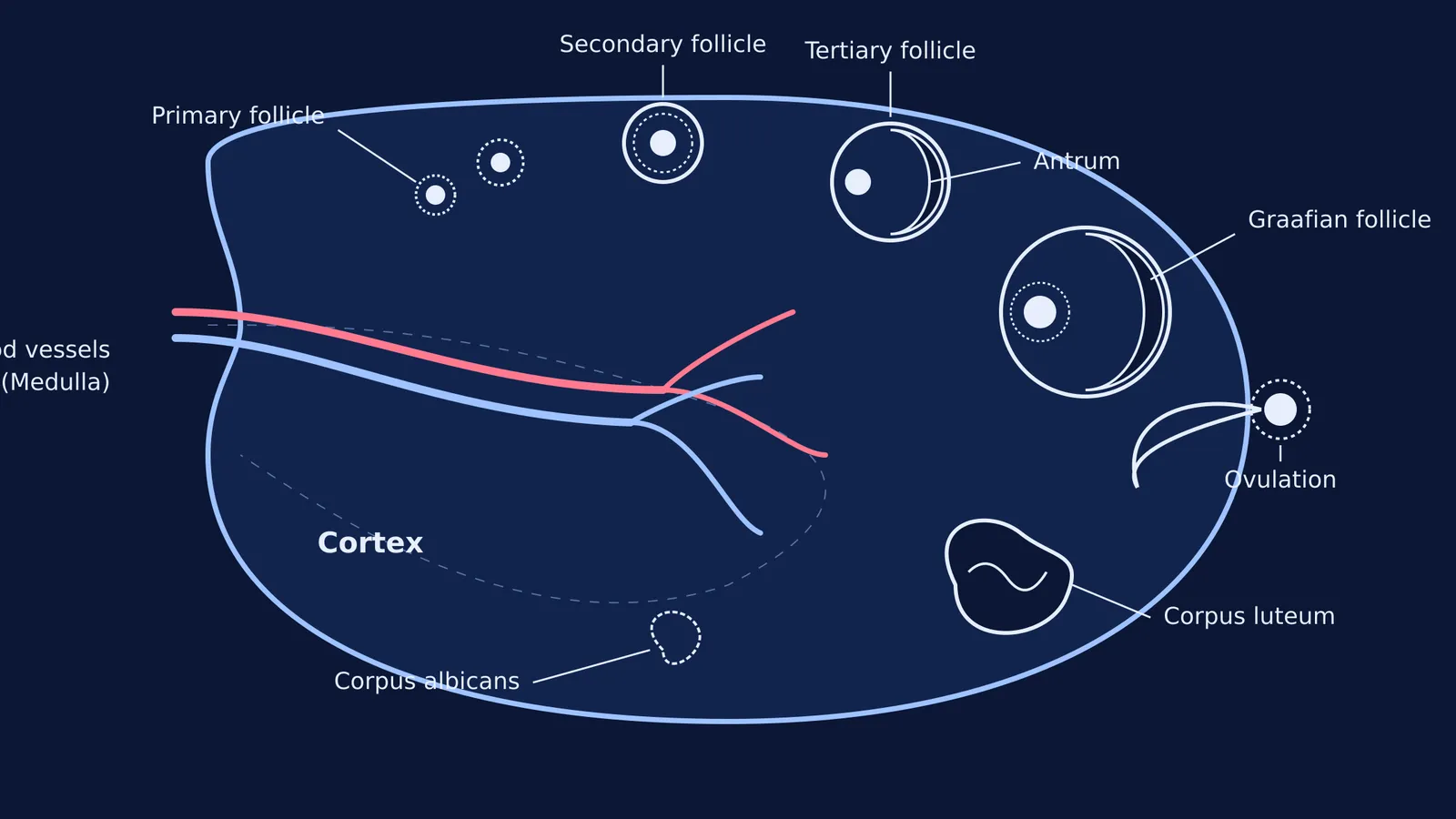
If you slice an ovary open, the tissue inside (the stroma) is divided into two distinct zones:
The ovarian medulla: The inner core. It is a highly vascular tissue, meaning it contains a rich network of blood vessels, lymphatic vessels and nerves that enter through the hilum.
The ovarian cortex: The outer region. This is where the action happens. The cortex houses the ovarian follicles at various stages of development.
Because the structures develop over time, a standard sectional view diagram displays all the stages of a follicle in a single continuous circle, almost like a time-lapse photograph captured in one frame.
What are the stages of a developing follicle?
At puberty, a young woman has around 200,000 to 300,000 follicles available in her ovaries. During each menstrual cycle, a few are selected to grow, progressing through the following stages visible in the cortex:
Primary follicle: A primary oocyte surrounded by a single layer of granulosa cells.
Secondary follicle: The follicle grows, adding more layers of granulosa cells and a new outer covering called the theca.
Tertiary follicle: This stage is easily identified in diagrams by the presence of a fluid-filled cavity called the antrum. The theca layer also organises into an inner and outer layer.
Graafian follicle: This is the fully mature follicle, named after the Dutch anatomist Regnier de Graaf. It has a large antrum and contains the secondary oocyte, ready for release.
If you struggle to recall the difference between these stages during your revision, the Fermi Concept Library breaks down the entire process of oogenesis into clear, logical steps.
What happens after ovulation?
Once the ovum is released into the fallopian tube, the story of the ruptured follicle continues:
Corpus luteum: The remaining granulosa and theca cells reorganise into a temporary endocrine gland called the corpus luteum (Latin for "yellow body"). It secretes large amounts of progesterone, which is essential to prepare the uterus for pregnancy.
Corpus albicans: If fertilisation does not occur, the corpus luteum degenerates, leaving behind a white scar tissue known as the corpus albicans.
When studying how these hormones interact with the rest of the reproductive cycle, Fermi's AI tutor can work out what you already know and build a plan for the session, ensuring you understand the feedback loops between the ovary and the pituitary gland.
How do you draw the ovary diagram?
If you need to draw this in a biology exam, follow this sequence to ensure you do not miss any labels:
[ ] Draw the boundary: Sketch an oval shape with a slight indentation (the hilum) on one side.
[ ] Add the blood vessels: Draw arteries and veins entering through the hilum into the central medulla.
[ ] Place the follicles in the cortex: Draw the progression of follicles in a sequence near the outer edge. Start small (primary), make them larger (secondary), and add a crescent-shaped space for the antrum (tertiary).
[ ] The Graafian follicle and ovum: Draw the largest follicle right at the edge, and show it rupturing to release the circular ovum.
[ ] The yellow body: Draw the irregularly shaped corpus luteum, followed by the smaller, shrinking corpus albicans.
By tracing the lifecycle of a follicle around the cortex, you can easily reconstruct the entire diagram from memory.
*Read next:*


