

On this page
In the first few days after fertilisation, the single-celled embryo divides rapidly—typically every 12 to 24 hours—as it travels down the fallopian tube. This rapid division is unique because the embryo does not grow larger overall; it simply splits the original cellular material into smaller and smaller pieces. By the time it reaches the uterus after this journey, this cluster of cells must organise itself for a critical task: attaching securely to the uterine wall to continue its development.
Understanding how the embryo reshapes itself during this time is a staple of NEET-UG biology and human embryology. Here is how a solid ball of cells turns into the structured blastocyst.
What is a morula?
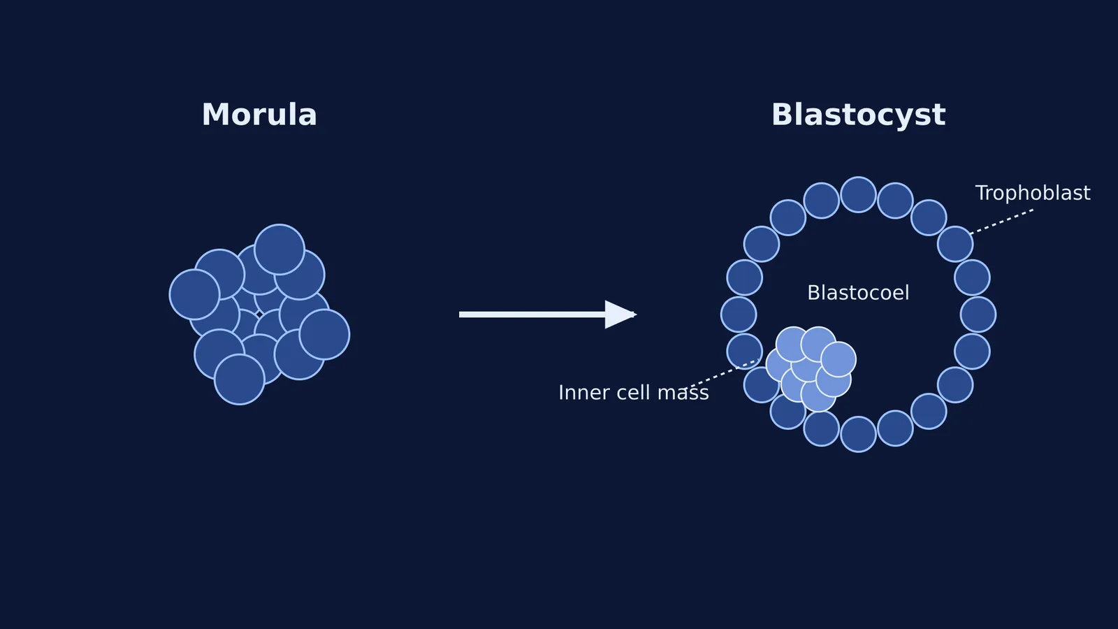
A morula is a solid, spherical ball of exactly 16 undifferentiated cells called blastomeres. At this stage, the embryo is still tumbling through the reproductive tract. The cells are tightly packed in a process called compaction, but they have not yet separated into different tissue types. Compaction is the crucial step that creates an internal population of cells, setting the stage for everything that follows. If you are tracking the sequence From fertilisation to implantation: the first week of pregnancy, the morula is the last stage before the embryo hollows out.
What is the difference between a morula and a blastocyst?
The key difference is structure: a morula is solid, while a blastocyst is hollow.
As the morula enters the uterus, it begins to take in fluid. This fluid pushes the cells around, creating a central cavity known as the blastocoel. Once this cavity forms, the embryo is officially a blastocyst.
Because embryology builds heavily on interrelated terms, missing one definition can make later stages confusing. If you have concept gaps around cleavage or cavitation, the Fermi Concept Library has every concept in biology, each with its own lesson and its place in the map.
What does the inner cell mass become?
When the blastocyst forms, its cells separate into two distinct groups. The inner cell mass is a cluster of cells pushed to one side of the cavity.
These are pluripotent cells, meaning they hold the potential to develop into almost any tissue type in the human body. Because of this remarkable flexibility, the inner cell mass will eventually give rise to the embryo proper—the actual fetus—as well as the associated membranes like the amnion that support it.
What is the role of the trophoblast?
The trophoblast is the outer layer of cells that forms the shell of the blastocyst.
While the inner cell mass focuses entirely on building the fetus, the trophoblast is entirely dedicated to survival and attachment. Its job is to burrow deep into the uterine wall during implantation. Once embedded, the trophoblast develops into the fetal portion of the placenta. This is the complex tissue that will eventually secrete essential hormones and coordinate nutrient exchange with maternal systems, a process later culminating in events like Parturition and lactation: the hormones behind birth and milk.
A checklist for the first week of development
When tackling embryology questions, verify that you can trace the fate of each structure:
Blastomeres: The individual cells that make up the early embryo.
Morula: The solid ball of 16 cells.
Blastocoel: The fluid cavity that turns the morula into a blastocyst.
Inner cell mass: The internal cluster that becomes the fetus.
Trophoblast: The outer layer that becomes the placenta.


