

On this page
Every eukaryotic cell that relies on aerobic respiration depends on mitochondria to generate adenosine triphosphate (ATP). Often introduced as the powerhouses of the cell, these organelles have a specialized compartmental architecture that directly drives their bioenergetic performance. For students preparing for NEET-UG and Class 11 CBSE examinations, understanding the structural division between the outer membrane, the inner membrane with its cristae, and the internal matrix is essential for mastering cellular respiration and cell biology.
What is the basic double-membrane structure of a mitochondrion?
A mitochondrion is an elongated, sausage-shaped or cylindrical organelle enclosed by two distinct concentric membranes. In curriculum standards aligned with CBSE Senior Secondary Biology and cell biology benchmarks (Alberts et al.), a typical mitochondrion measures between 0.2 µm and 1.0 µm in diameter (average 0.5 µm) and reaches a length of 1.0 µm to 4.1 µm.
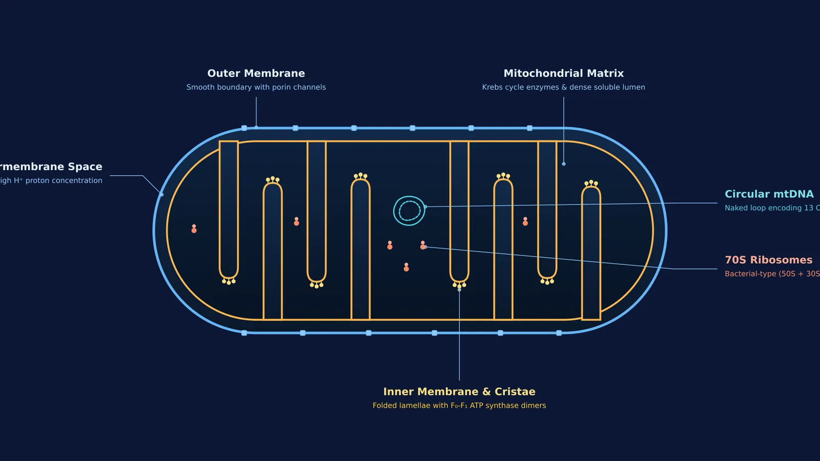
The two membranes partition the organelle into two aqueous compartments: the intermembrane space and the internal matrix. The outer membrane serves as a continuous, smooth boundary. It contains transmembrane proteins called porins, which form large aqueous channels that allow ions, nucleotides, and small metabolic substrates up to approximately 5,000 Daltons to diffuse freely (Alberts et al.).
The inner membrane lies immediately inside the outer membrane and exhibits strict selective permeability. Unlike the outer membrane, it is rich in the unique diphosphatidylglycerol lipid cardiolipin and nearly impermeable to ions and protons. This barrier ensures that protons pumped into the intermembrane space during electron transport cannot leak backward randomly, preserving the vital electrochemical potential difference required for ATP production. You can review how these boundaries contrast with single-membrane organelles in our guide to Cell: the unit of life for NEET.
How do cristae increase surface area for ATP synthesis?
The inner mitochondrial membrane does not form a smooth inner boundary; instead, it projects inward toward the matrix in numerous deep folds termed cristae (singular: crista). The fundamental biological function of cristae is to expand the inner membrane surface area, providing room for thousands of electron transport chain complexes and ATP synthase motors.
Structural investigations using electron cryotomography published in PNAS by Davies and colleagues (2012) revealed that ATP synthase molecules form V-shaped dimers with an angle of roughly 86° between monomers. These dimers assemble into long oligomeric ribbons that actively induce curvature along the inner membrane, stabilizing the sharp apex of each crista fold.
This geometric curvature serves an important bioenergetic purpose:
Electron transport complexes (Complexes I, III, and IV) pump hydrogen ions from the matrix into the narrow crista lumen.
The restricted volume of the crista lumen traps protons locally, raising the proton concentration relative to the bulk intermembrane space.
The high local proton concentration funnels through the catalytic F₀-F₁ ATP synthase particles localized along the crista ridges.
To trace how metabolic substrates feed into these respiratory assemblies, read our detailed breakdown of respiration in plants: how to count ATP from glycolysis to ETS.
What does the mitochondrial matrix contain?
Enclosed by the inner membrane is the inner compartment, which is filled with a dense, homogeneous, gel-like substance known as the mitochondrial matrix. The matrix contains the enzymatic machinery required for metabolic fuel breakdown alongside an autonomous genetic system.
Key biochemical constituents of the matrix include:
A single circular, double-stranded DNA molecule that lacks histone proteins, mirroring bacterial chromosomes.
70S ribosomes, consisting of 50S and 30S subunits, which differ from the larger 80S ribosomes found in the eukaryotic cytoplasm.
Transfer RNA (tRNA) molecules, messenger RNA transcripts, and the full set of enzymes needed to translate proteins.
Soluble enzymes catalyzing the citric acid (Krebs) cycle, pyruvate decarboxylation, and fatty acid beta-oxidation.
Because mitochondria contain their own DNA and 70S ribosomes, they are classified as semi-autonomous organelles. As established by Anderson and colleagues (1981), human mitochondrial DNA encodes approximately 13 core polypeptide subunits of the respiratory complexes locally, while the remaining mitochondrial proteins are translated on cytoplasmic ribosomes and imported from the cytosol.
How do mitochondria divide by fission?
Mitochondria cannot be generated from scratch by the cell. Instead, new mitochondria arise exclusively through binary fission of pre-existing organelles.
Research published in Science by Friedman and colleagues (2011) established that tubules of the endoplasmic reticulum wrap around the mitochondrion to initiate initial constriction, after which dynamin-related GTPase rings (Drp1 in humans) assemble at the contact site to pinch the inner and outer membranes apart.
This division process closely parallels bacterial reproduction. In 1967, Lynn Sagan (Margulis)90079-3) synthesized these structural clues into the endosymbiotic theory, proposing that mitochondria originated when primitive eukaryotic cells engulfed aerobic alpha-proteobacteria. The presence of circular DNA, 70S ribosomes, binary fission, and an inner membrane with bacterial-type lipids provides compelling proof of this evolutionary history. You can explore this milestone in our feature on Lynn Margulis and the bacteria inside your cells.
To explore how cellular anatomy fits into broader biological systems, the Fermi Concept Library maps every concept across biology, giving each topic its own lesson and clear place in the map.
How should you revise mitochondrial structure for NEET?
When reviewing mitochondrial anatomy for examinations aligned with the CBSE Senior Secondary Biology curriculum, examiners test whether you can distinguish between membrane compartments, enzymatic localizations, and endosymbiotic characteristics.
Use this four-point diagnostic checklist during your revisions:
Outer membrane: Smooth and permeable to small molecules below 5,000 Daltons via porin channels (Alberts et al.).
Inner membrane: Infolded into cristae to maximize surface area; contains cardiolipin, electron transport complexes I through IV, and F₀-F₁ ATP synthase particles.
Matrix: Dense inner compartment housing circular DNA, 70S ribosomes, RNA, and Krebs cycle enzymes (with succinate dehydrogenase as the sole membrane-bound exception).
Reproduction and genetics: Replicates strictly by fission and exhibits maternal cytoplasmic inheritance in humans.
When solving multi-statement questions or practice tests on organelle structure, the Fermi Problem Library provides worked problems with a step-by-step solver that explains the reasoning rather than printing an answer.


