

On this page
The human mammary gland is an accessory reproductive organ classified anatomically as a modified apocrine sweat gland. Resting over the pectoralis major muscle of the anterior thoracic wall, its functional parenchyma is supported by fibrous bands called Cooper's ligaments and variable amounts of adipose tissue. According to StatPearls anatomical guidelines, each breast contains 15 to 20 mammary lobes arranged radially around the nipple-areolar complex. Each individual lobe functions as an independent glandular unit subdivided into smaller lobules, which house the microscopic secretory units known as alveoli.
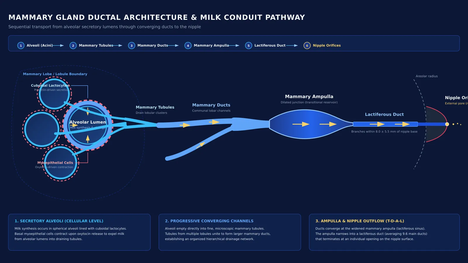
At the cellular level, the alveoli are spherical acini lined with a single layer of milk-secreting cuboidal epithelial cells called lactocytes. These secretory cells synthesize proteins, lactose, and lipid droplets, discharging them directly into the central cavity, or lumen, of each alveolus. As shown by Brisken and Rajaram (2006), prolactin drives this secretory differentiation of the alveolar epithelium during late pregnancy and postpartum lactation. Surrounding the basal surface of the epithelial cells is a web of contractile myoepithelial cells, which form a responsive basket-like scaffolding around each alveolus.
How does milk flow from alveoli to the nipple?
Milk does not seep passively across the breast; it travels through an organized conduit tree that narrows, merges, widens, and discharges in an exact sequence:
Alveolar lumens: Milk is synthesized by epithelial lactocytes and collects within the hollow centre of the alveoli.
Mammary tubules: The alveoli open directly into fine, microscopic tubes called mammary tubules that drain each lobular cluster.
Mammary ducts: Tubules from different lobules within each lobe converge to build a larger communal channel called a mammary duct.
Mammary ampulla: Multiple mammary ducts unite into an expanded, wider section called the mammary ampulla, which serves as a transitional reservoir.
Lactiferous ducts: The ampulla narrows into a robust lactiferous duct that directs milk through the subareolar zone toward the surface.
Nipple orifices: Each lactiferous duct terminates at an individual opening, allowing milk to exit when suckling creates negative external pressure.
For decades, standard medical diagrams depicted large bulbous sacs storing milk directly behind the areola. However, high-resolution ultrasound imaging published by Ramsay and colleagues (2005) across 21 lactating women demonstrated that milk ducts are superficial, easily compressible conduits that branch within 8.0 ± 5.5 mm of the nipple base, with an average of 9.6 ± 2.9 main duct systems in the left breast and 9.2 ± 2.9 in the right breast. Rather than static reservoirs, these ducts function primarily as dynamic conduits. Forward transport relies on a neuroendocrine reflex reviewed by Walter, Abele, and Plappert (2021): infant suckling stimulates sensory afferents, prompting the posterior pituitary to pulse oxytocin into the bloodstream, which causes myoepithelial cells around the alveoli to contract and propel milk outward.
Why do NEET questions focus on the ductal sequence?
In competitive biology exams like NEET-UG, questions about the mammary gland test anatomical hierarchy rather than rote definitions. Test-makers frequently target the transition zones where students confuse microscopic branches with macro-level conduits. The most common errors include reversing the order of tubules and ducts, confusing the ampulla with the lactiferous duct, or misidentifying the cell types responsible for milk production versus milk ejection.
Reviewing past patterns in human reproduction question formats for NEET shows that sequence arrangement problems appear regularly. Understanding how cellular secretion connects with the larger hormonal triggers behind parturition and lactation helps you answer both direct recall questions and assertion-reason items. When you need to review the hierarchy from cell to organ, the Fermi Concept Library covers every concept in biology with its own lesson and its place in the map.
How can you verify the milk pathway under exam pressure?
To avoid second-guessing during timed tests, apply a four-step diagnostic check to any sequence question:
Origin check: Milk synthesis begins exclusively in the alveolar lumen, never in connective stroma or adipose tissue.
Calibre check: "Tubule" denotes a tiny conduit, so mammary tubules must precede the larger mammary ducts.
Dilation check: The ampulla is a wide junction point where several mammary ducts pool before entering the terminal conduit.
Exit check: The lactiferous duct is the final passage connecting the ampulla to the external pore on the nipple.
Practicing sequence problems with active recall prevents exam-room confusion between neuroendocrine triggers and physical conduits. The broader regulatory system relies on reciprocal endocrine feedback loops where prolactin maintains synthesis while oxytocin regulates ejection. For targeted practice on sequence questions, the Fermi Problem Library has worked problems with a step-by-step solver that explains the reasoning rather than printing an answer. If you find yourself stuck distinguishing structural anatomy from hormonal control, Fermi's AI tutor can work out what you already know and build a plan for the session, drawing the figure and checking understanding as you go.


